• 文献检索
  • 文档翻译
  • 深度研究
  • 学术资讯
  • Suppr Zotero 插件Zotero 插件
  • 邀请有礼
  • 套餐&价格
  • 历史记录
应用&插件
Suppr Zotero 插件Zotero 插件浏览器插件Mac 客户端Windows 客户端微信小程序
定价
高级版会员购买积分包购买API积分包
服务
文献检索文档翻译深度研究API 文档MCP 服务
关于我们
关于 Suppr公司介绍联系我们用户协议隐私条款
关注我们

Suppr 超能文献

核心技术专利:CN118964589B侵权必究
粤ICP备2023148730 号-1Suppr @ 2026

文献检索

告别复杂PubMed语法,用中文像聊天一样搜索,搜遍4000万医学文献。AI智能推荐,让科研检索更轻松。

立即免费搜索

文件翻译

保留排版,准确专业,支持PDF/Word/PPT等文件格式,支持 12+语言互译。

免费翻译文档

深度研究

AI帮你快速写综述,25分钟生成高质量综述,智能提取关键信息,辅助科研写作。

立即免费体验

丹麦高街配镜师环境下视网膜图像中血管形态计量的自动分析。

Automated analysis of vessel morphometry in retinal images from a Danish high street optician setting.

机构信息

Department of Drug Design and Pharmacology, University of Copenhagen, Copenhagen, Denmark.

School of Computer Science and Mathematics, Kingston University, Surrey, United Kingdom.

出版信息

PLoS One. 2023 Aug 24;18(8):e0290278. doi: 10.1371/journal.pone.0290278. eCollection 2023.

DOI:10.1371/journal.pone.0290278
PMID:37616264
原文链接:https://pmc.ncbi.nlm.nih.gov/articles/PMC10449151/
Abstract

PURPOSE

To evaluate the test performance of the QUARTZ (QUantitative Analysis of Retinal vessel Topology and siZe) software in detecting retinal features from retinal images captured by health care professionals in a Danish high street optician chain, compared with test performance from other large population studies (i.e., UK Biobank) where retinal images were captured by non-experts.

METHOD

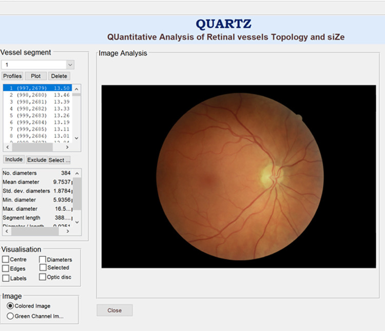
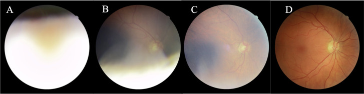
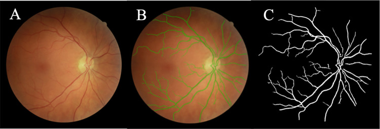
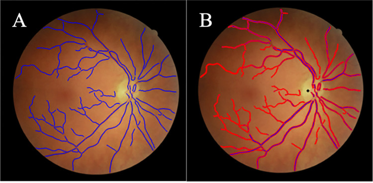
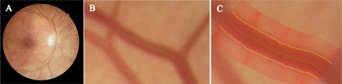
The dataset FOREVERP (Finding Ophthalmic Risk and Evaluating the Value of Eye exams and their predictive Reliability, Pilot) contains retinal images obtained from a Danish high street optician chain. The QUARTZ algorithm utilizes both image processing and machine learning methods to determine retinal image quality, vessel segmentation, vessel width, vessel classification (arterioles or venules), and optic disc localization. Outcomes were evaluated by metrics including sensitivity, specificity, and accuracy and compared to human expert ground truths.

RESULTS

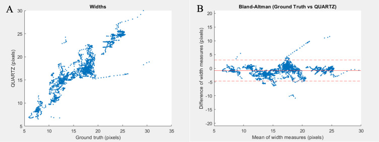
QUARTZ's performance was evaluated on a subset of 3,682 images from the FOREVERP database. 80.55% of the FOREVERP images were labelled as being of adequate quality compared to 71.53% of UK Biobank images, with a vessel segmentation sensitivity of 74.64% and specificity of 98.41% (FOREVERP) compared with a sensitivity of 69.12% and specificity of 98.88% (UK Biobank). The mean (± standard deviation) vessel width of the ground truth was 16.21 (4.73) pixels compared to that predicted by QUARTZ of 17.01 (4.49) pixels, resulting in a difference of -0.8 (1.96) pixels. The differences were stable across a range of vessels. The detection rate for optic disc localisation was similar for the two datasets.

CONCLUSION

QUARTZ showed high performance when evaluated on the FOREVERP dataset, and demonstrated robustness across datasets, providing validity to direct comparisons and pooling of retinal feature measures across data sources.

摘要

目的

评估 QUARTZ(视网膜血管拓扑和大小的定量分析)软件在检测丹麦高街配镜师连锁店中医疗保健专业人员拍摄的视网膜图像中的视网膜特征的测试性能,与其他大型人群研究(即英国生物库)的测试性能进行比较,这些研究中的视网膜图像是由非专业人员拍摄的。

方法

FOREVERP(发现眼科风险并评估眼部检查及其预测可靠性、试点)数据集包含从丹麦高街配镜师连锁店获得的视网膜图像。QUARTZ 算法利用图像处理和机器学习方法来确定视网膜图像质量、血管分割、血管宽度、血管分类(小动脉或小静脉)和视盘定位。通过包括敏感性、特异性和准确性在内的指标来评估结果,并与人类专家的真实情况进行比较。

结果

QUARTZ 的性能在 FOREVERP 数据库的一个子集中进行了评估。与 71.53%的英国生物库图像相比,FOREVERP 图像中有 80.55%被标记为具有足够质量,血管分割敏感性为 74.64%,特异性为 98.41%(FOREVERP),而敏感性为 69.12%,特异性为 98.88%(英国生物库)。地面真实的平均(±标准偏差)血管宽度为 16.21(4.73)像素,而 QUARTZ 预测的血管宽度为 17.01(4.49)像素,差值为-0.8(1.96)像素。差异在各种血管中均保持稳定。两个数据集的视盘定位检测率相似。

结论

QUARTZ 在 FOREVERP 数据集上的评估表现出了很高的性能,并且在数据集之间表现出了稳健性,为直接比较和跨数据源的视网膜特征测量值的汇总提供了有效性。

https://cdn.ncbi.nlm.nih.gov/pmc/blobs/3515/10449151/6eb1685cba94/pone.0290278.g006.jpg
https://cdn.ncbi.nlm.nih.gov/pmc/blobs/3515/10449151/ed59519fa79c/pone.0290278.g001.jpg
https://cdn.ncbi.nlm.nih.gov/pmc/blobs/3515/10449151/eae5b691753d/pone.0290278.g002.jpg
https://cdn.ncbi.nlm.nih.gov/pmc/blobs/3515/10449151/adde84eb88f3/pone.0290278.g003.jpg
https://cdn.ncbi.nlm.nih.gov/pmc/blobs/3515/10449151/e53eb61c8cf6/pone.0290278.g004.jpg
https://cdn.ncbi.nlm.nih.gov/pmc/blobs/3515/10449151/82490d3a80e2/pone.0290278.g005.jpg
https://cdn.ncbi.nlm.nih.gov/pmc/blobs/3515/10449151/6eb1685cba94/pone.0290278.g006.jpg

相似文献

1
Automated analysis of vessel morphometry in retinal images from a Danish high street optician setting.丹麦高街配镜师环境下视网膜图像中血管形态计量的自动分析。
PLoS One. 2023 Aug 24;18(8):e0290278. doi: 10.1371/journal.pone.0290278. eCollection 2023.
2
Automated arteriole and venule classification using deep learning for retinal images from the UK Biobank cohort.使用深度学习对 UK Biobank 队列的视网膜图像进行自动微动脉和微静脉分类。
Comput Biol Med. 2017 Nov 1;90:23-32. doi: 10.1016/j.compbiomed.2017.09.005. Epub 2017 Sep 8.
3
Automated retinal image quality assessment on the UK Biobank dataset for epidemiological studies.用于流行病学研究的英国生物银行数据集上的视网膜图像质量自动评估。
Comput Biol Med. 2016 Apr 1;71:67-76. doi: 10.1016/j.compbiomed.2016.01.027. Epub 2016 Feb 6.
4
MSGANet-RAV: A multiscale guided attention network for artery-vein segmentation and classification from optic disc and retinal images.MSGANet-RAV:一种用于从视盘和视网膜图像中进行动脉-静脉分割和分类的多尺度引导注意力网络。
J Optom. 2022;15 Suppl 1(Suppl 1):S58-S69. doi: 10.1016/j.optom.2022.11.001. Epub 2022 Nov 14.
5
A new robust method for blood vessel segmentation in retinal fundus images based on weighted line detector and hidden Markov model.基于加权线检测器和隐马尔可夫模型的视网膜眼底图像血管分割新方法。
Comput Methods Programs Biomed. 2020 Apr;187:105231. doi: 10.1016/j.cmpb.2019.105231. Epub 2019 Nov 23.
6
A Deep-Learning Approach for Automated OCT En-Face Retinal Vessel Segmentation in Cases of Optic Disc Swelling Using Multiple En-Face Images as Input.一种基于深度学习的方法,用于使用多张 OCT 眼前节图像作为输入,对视盘肿胀病例进行 OCT 自动眼前节视网膜血管分割。
Transl Vis Sci Technol. 2020 Mar 24;9(2):17. doi: 10.1167/tvst.9.2.17. eCollection 2020 Mar.
7
Two-stage framework for optic disc localization and glaucoma classification in retinal fundus images using deep learning.基于深度学习的眼底图像视盘定位和青光眼分类的两阶段框架。
BMC Med Inform Decis Mak. 2019 Jul 17;19(1):136. doi: 10.1186/s12911-019-0842-8.
8
Segmentation of retinal blood vessels by a novel hybrid technique- Principal Component Analysis (PCA) and Contrast Limited Adaptive Histogram Equalization (CLAHE).基于主成分分析(PCA)和对比度受限自适应直方图均衡化(CLAHE)的新型混合技术对视网膜血管的分割。
Microvasc Res. 2023 Jul;148:104477. doi: 10.1016/j.mvr.2023.104477. Epub 2023 Feb 4.
9
Finding Ophthalmic Risk and Evaluating the Value of Eye exams and their predictive Reliability (FOREVER)-A cohort study in a Danish high street optician setting: Design and methodology.寻找眼科风险并评估眼科检查的价值及其预测可靠性(FOREVER)-丹麦高街配镜师环境中的队列研究:设计与方法。
Acta Ophthalmol. 2024 Feb;102(1):80-90. doi: 10.1111/aos.15693. Epub 2023 May 4.
10
Reliability of vessel diameter measurements with a retinal oximeter.视网膜血氧计测量血管直径的可靠性。
Graefes Arch Clin Exp Ophthalmol. 2011 Sep;249(9):1311-7. doi: 10.1007/s00417-011-1680-2. Epub 2011 Apr 16.

引用本文的文献

1
Longitudinal associations of retinal vessel morphology with intraocular pressure and blood pressure at follow-up visit-Findings from a Danish eye and vision cohort, Project FOREVER.随访时视网膜血管形态与眼压和血压的纵向关联——来自丹麦眼睛与视力队列研究“永恒计划”的发现
Acta Ophthalmol. 2025 Feb;103(1):33-42. doi: 10.1111/aos.16737. Epub 2024 Jul 2.

本文引用的文献

1
Finding Ophthalmic Risk and Evaluating the Value of Eye exams and their predictive Reliability (FOREVER)-A cohort study in a Danish high street optician setting: Design and methodology.寻找眼科风险并评估眼科检查的价值及其预测可靠性(FOREVER)-丹麦高街配镜师环境中的队列研究:设计与方法。
Acta Ophthalmol. 2024 Feb;102(1):80-90. doi: 10.1111/aos.15693. Epub 2023 May 4.
2
Artificial intelligence-enabled retinal vasculometry for prediction of circulatory mortality, myocardial infarction and stroke.人工智能赋能的视网膜血管测量学预测循环死亡率、心肌梗死和中风。
Br J Ophthalmol. 2022 Dec;106(12):1722-1729. doi: 10.1136/bjo-2022-321842. Epub 2022 Oct 4.
3
Machine-Learning-Based Disease Diagnosis: A Comprehensive Review.
基于机器学习的疾病诊断:全面综述
Healthcare (Basel). 2022 Mar 15;10(3):541. doi: 10.3390/healthcare10030541.
4
Applications of interpretability in deep learning models for ophthalmology.深度学习模型在眼科领域的可解释性应用。
Curr Opin Ophthalmol. 2021 Sep 1;32(5):452-458. doi: 10.1097/ICU.0000000000000780.
5
Predicting the risk of developing diabetic retinopathy using deep learning.利用深度学习预测糖尿病视网膜病变的风险。
Lancet Digit Health. 2021 Jan;3(1):e10-e19. doi: 10.1016/S2589-7500(20)30250-8. Epub 2020 Nov 26.
6
The Right Direction Needed to Develop White-Box Deep Learning in Radiology, Pathology, and Ophthalmology: A Short Review.放射学、病理学和眼科领域发展白盒深度学习所需的正确方向:简要综述
Front Robot AI. 2019 Apr 16;6:24. doi: 10.3389/frobt.2019.00024. eCollection 2019.
7
A review of machine learning methods for retinal blood vessel segmentation and artery/vein classification.机器学习方法在视网膜血管分割和动静脉分类中的研究进展综述。
Med Image Anal. 2021 Feb;68:101905. doi: 10.1016/j.media.2020.101905. Epub 2020 Nov 17.
8
A Hybrid Unsupervised Approach for Retinal Vessel Segmentation.一种混合无监督的视网膜血管分割方法。
Biomed Res Int. 2020 Dec 10;2020:8365783. doi: 10.1155/2020/8365783. eCollection 2020.
9
Robust and Interpretable Convolutional Neural Networks to Detect Glaucoma in Optical Coherence Tomography Images.稳健且可解释的卷积神经网络,用于检测光学相干断层扫描图像中的青光眼。
IEEE Trans Biomed Eng. 2021 Aug;68(8):2456-2466. doi: 10.1109/TBME.2020.3043215. Epub 2021 Jul 19.
10
Retinal blood flow in critical illness and systemic disease: a review.危重症和全身性疾病中的视网膜血流:综述
Ann Intensive Care. 2020 Nov 12;10(1):152. doi: 10.1186/s13613-020-00768-3.