• 文献检索
  • 文档翻译
  • 深度研究
  • 学术资讯
  • Suppr Zotero 插件Zotero 插件
  • 邀请有礼
  • 套餐&价格
  • 历史记录
应用&插件
Suppr Zotero 插件Zotero 插件浏览器插件Mac 客户端Windows 客户端微信小程序
定价
高级版会员购买积分包购买API积分包
服务
文献检索文档翻译深度研究API 文档MCP 服务
关于我们
关于 Suppr公司介绍联系我们用户协议隐私条款
关注我们

Suppr 超能文献

核心技术专利:CN118964589B侵权必究
粤ICP备2023148730 号-1Suppr @ 2026

文献检索

告别复杂PubMed语法,用中文像聊天一样搜索,搜遍4000万医学文献。AI智能推荐,让科研检索更轻松。

立即免费搜索

文件翻译

保留排版,准确专业,支持PDF/Word/PPT等文件格式,支持 12+语言互译。

免费翻译文档

深度研究

AI帮你快速写综述,25分钟生成高质量综述,智能提取关键信息,辅助科研写作。

立即免费体验

头对头比较增强 CT、68Ga-PSMA-11 PET/CT 和 18F-FDG PET/CT 识别肾透明细胞癌不良病理:一项前瞻性研究。

Head-to-head comparisons of enhanced CT, 68Ga-PSMA-11 PET/CT and 18F-FDG PET/CT in identifying adverse pathology of clear-cell renal cell carcinoma: a prospective study.

机构信息

Department of Urology, Urology Research Institute, The First Affiliated Hospital of Fujian Medical University, Fuzhou, China.

Department of Urology, National Region Medical center, Binhai Campus of the First Affiliated Hospital, Fujian Medical University, Fuzhou, China.

出版信息

Int Braz J Urol. 2023 Nov-Dec;49(6):716-731. doi: 10.1590/S1677-5538.IBJU.2023.0312.

DOI:10.1590/S1677-5538.IBJU.2023.0312
PMID:37624658
原文链接:https://pmc.ncbi.nlm.nih.gov/articles/PMC10947621/
Abstract

OBJECTIVES

Accurate preoperative prediction of adverse pathology is crucial for treatment planning of renal cell carcinoma (RCC). Previous studies have emphasized the potential of prostate-specific membrane antigen positron emission tomography / computed tomography (PSMA PET/CT) in differentiating between benign and malignant localized renal tumors. However, there is a scarcity of case reports elucidating the identification of aggressive pathological features using PET/CT. Our study was designed to prospectively compare the diagnostic value of enhanced CT, 68Ga-PSMA-11 and 18F-fluorodeoxyglucose (18F-FDG) PET/CT in clear-cell renal cell carcinoma (ccRCC) with necrosis or sarcomatoid or rhabdoid differentiation.

MATERIALS AND METHODS

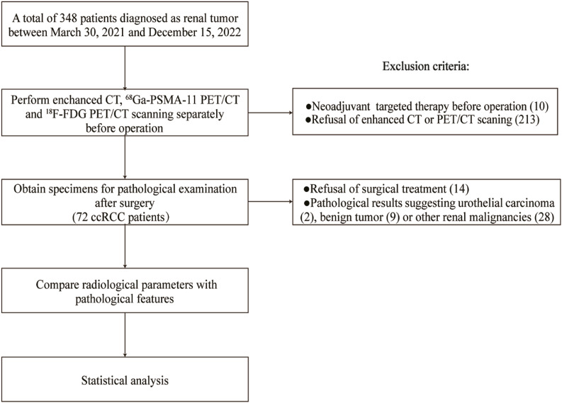
A prospective case series of patients with a newly diagnosed renal mass who underwent enhanced CT, 68Ga-PSMA-11 and 18F-FDG PET/CT within 30 days prior to nephrectomy was included. Complete preoperative and postoperative clinicopathological data were recorded. Patients who received neoadjuvant targeted therapy, declined enhanced CT or PET/CT scanning, refused surgical treatment or had non-ccRCC pathological indications were excluded. Radiological parameters were compared within subgroups of pathological characteristics. Bonferroni corrections were used to adjust for multiple testing and statistical significance was set at a p-value less than 0.017.

RESULTS

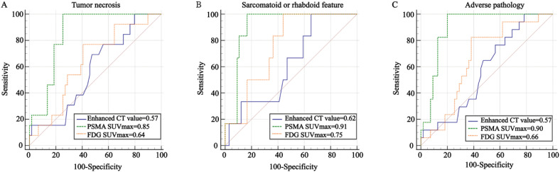
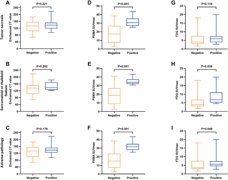
Seventy-two patients were available for the final analysis. Enhanced CT demonstrated poor performance in identifying necrosis, sarcomatoid or rhabdoid differentiation and adverse pathology (all P > 0.05). The maximum standardized uptake value (SUVmax) of 68Ga-PSMA-11 PET/CT was more effective than 18F-FDG PET/CT in identifying tumor necrosis and adverse pathology, with an area under the curve (AUC) of 0.85 (cutoff value=25.26, p<0.001; Delong test z=2.709, p=0.007) for tumor necrosis and AUC of 0.90 (cutoff value=25.26, p<0.001; Delong test z=3.433, p<0.001) for adverse pathology. However, no significant statistical difference was found between 68Ga-PSMA-11 and 18F-FDG PET/CT in predicting sarcomatoid or rhabdoid feature (AUC of 0.91 vs.0.75, Delong test z=1.998, p=0.046). Subgroup analyses based on age, sex, tumor location, maximal diameter, stage and WHO/ISUP grade demonstrated that 68Ga-PSMA-11 PET/CT SUVmax had a significant predictive value for adverse pathology. Enhanced CT value and SUVmax demonstrated strong reliability [intraclass correlation coefficient (ICC) > 0.80], indicating a robust correlation.

CONCLUSIONS

68Ga-PSMA-11 PET/CT demonstrates distinct advantages in identifying aggressive pathological features of primary ccRCC when compared to enhanced CT and 18F-FDG PET/CT. Further research and assessment are warranted to fully establish the clinical utility of 68Ga-PSMA-11 PET/CT in ccRCC.

摘要

目的

准确预测术前不良病理对肾细胞癌(RCC)的治疗计划至关重要。先前的研究强调了前列腺特异性膜抗原正电子发射断层扫描/计算机断层扫描(PSMA PET/CT)在区分良性和恶性局限性肾肿瘤方面的潜力。然而,很少有病例报告阐明了使用 PET/CT 识别侵袭性病理特征的情况。我们的研究旨在前瞻性比较增强 CT、68Ga-PSMA-11 和 18F-氟脱氧葡萄糖(18F-FDG)PET/CT 在伴有坏死或肉瘤样或横纹肌样分化的透明细胞肾细胞癌(ccRCC)中的诊断价值。

材料和方法

纳入了一组接受新诊断的肾肿块患者的前瞻性病例系列,这些患者在肾切除术 30 天内接受了增强 CT、68Ga-PSMA-11 和 18F-FDG PET/CT 检查。记录了完整的术前和术后临床病理数据。排除了接受新辅助靶向治疗、拒绝增强 CT 或 PET/CT 扫描、拒绝手术治疗或具有非 ccRCC 病理指征的患者。在病理特征的亚组内比较了影像学参数。使用 Bonferroni 校正进行多重检验调整,统计学意义设定为 p 值小于 0.017。

结果

72 例患者最终纳入分析。增强 CT 在识别坏死、肉瘤样或横纹肌样分化和不良病理方面表现不佳(均 P > 0.05)。68Ga-PSMA-11 PET/CT 的最大标准化摄取值(SUVmax)在识别肿瘤坏死和不良病理方面优于 18F-FDG PET/CT,曲线下面积(AUC)为 0.85(截断值=25.26,p<0.001;Delong 检验 z=2.709,p=0.007)用于肿瘤坏死和 AUC 为 0.90(截断值=25.26,p<0.001;Delong 检验 z=3.433,p<0.001)用于不良病理。然而,68Ga-PSMA-11 和 18F-FDG PET/CT 在预测肉瘤样或横纹肌样特征方面没有显著的统计学差异(AUC 为 0.91 与 0.75,Delong 检验 z=1.998,p=0.046)。基于年龄、性别、肿瘤位置、最大直径、分期和世卫组织/国际泌尿病理学会分级的亚组分析表明,68Ga-PSMA-11 PET/CT SUVmax 对不良病理具有显著的预测价值。增强 CT 值和 SUVmax 表现出很强的可靠性[组内相关系数(ICC)>0.80],表明相关性很强。

结论

与增强 CT 和 18F-FDG PET/CT 相比,68Ga-PSMA-11 PET/CT 在识别原发性 ccRCC 的侵袭性病理特征方面具有明显优势。需要进一步研究和评估,以充分确定 68Ga-PSMA-11 PET/CT 在 ccRCC 中的临床应用价值。

https://cdn.ncbi.nlm.nih.gov/pmc/blobs/b645/10947621/8c8b1e036c7a/1677-6119-ibju-49-06-0716-gf04.jpg
https://cdn.ncbi.nlm.nih.gov/pmc/blobs/b645/10947621/8ad7c9b2dd07/1677-6119-ibju-49-06-0716-gf01.jpg
https://cdn.ncbi.nlm.nih.gov/pmc/blobs/b645/10947621/701eb0dca6ba/1677-6119-ibju-49-06-0716-gf02.jpg
https://cdn.ncbi.nlm.nih.gov/pmc/blobs/b645/10947621/924a4529cfec/1677-6119-ibju-49-06-0716-gf03.jpg
https://cdn.ncbi.nlm.nih.gov/pmc/blobs/b645/10947621/8c8b1e036c7a/1677-6119-ibju-49-06-0716-gf04.jpg

相似文献

1
Head-to-head comparisons of enhanced CT, 68Ga-PSMA-11 PET/CT and 18F-FDG PET/CT in identifying adverse pathology of clear-cell renal cell carcinoma: a prospective study.头对头比较增强 CT、68Ga-PSMA-11 PET/CT 和 18F-FDG PET/CT 识别肾透明细胞癌不良病理:一项前瞻性研究。
Int Braz J Urol. 2023 Nov-Dec;49(6):716-731. doi: 10.1590/S1677-5538.IBJU.2023.0312.
2
Comprehensive evaluation of Ga-PSMA-11 PET/CT parameters for discriminating pathological characteristics in primary clear-cell renal cell carcinoma.全面评估 Ga-PSMA-11 PET/CT 参数在原发性透明细胞肾细胞癌病理特征鉴别中的价值。
Eur J Nucl Med Mol Imaging. 2021 Feb;48(2):561-569. doi: 10.1007/s00259-020-04916-6. Epub 2020 Jul 4.
3
[Ga]Ga‑LNC1007 PET/CT in the evaluation of renal cell carcinoma: comparison with 2-[F]FDG/[Ga]Ga-PSMA PET/CT.[镓]镓‑LNC1007 PET/CT 在肾细胞癌评估中的应用:与 2-[F]FDG/[镓]镓-PSMA PET/CT 的比较。
Eur J Nucl Med Mol Imaging. 2024 Jan;51(2):535-547. doi: 10.1007/s00259-023-06436-5. Epub 2023 Sep 20.
4
FDG PET/CT and CT Findings of Renal Cell Carcinoma With Sarcomatoid Differentiation.具有肉瘤样分化的肾细胞癌的 FDG PET/CT 和 CT 表现。
AJR Am J Roentgenol. 2020 Sep;215(3):645-651. doi: 10.2214/AJR.19.22467. Epub 2020 Jul 1.
5
Head-to-head comparison of [Ga]Ga-P16-093 and 2-[F]FDG PET/CT in patients with clear cell renal cell carcinoma: a pilot study.[镓]Ga-P16-093与2-[氟]FDG PET/CT在透明细胞肾细胞癌患者中的头对头比较:一项初步研究。
Eur J Nucl Med Mol Imaging. 2023 Apr;50(5):1499-1509. doi: 10.1007/s00259-022-06101-3. Epub 2023 Jan 5.
6
Potential role of Ga-PSMA PET/CT in metastatic renal cell cancer: A prospective study.镓-PSMA PET/CT 在转移性肾细胞癌中的潜在作用:一项前瞻性研究。
Eur J Radiol. 2024 Jan;170:111218. doi: 10.1016/j.ejrad.2023.111218. Epub 2023 Nov 23.
7
Diagnostic performance and prognostic value of preoperative F-FDG PET/CT in renal cell carcinoma patients with venous tumor thrombus.术前 F-FDG PET/CT 对伴有静脉瘤栓的肾细胞癌患者的诊断性能和预后价值。
Cancer Imaging. 2022 Nov 26;22(1):65. doi: 10.1186/s40644-022-00502-1.
8
More advantages in detecting bone and soft tissue metastases from prostate cancer using F-PSMA PET/CT.使用F-PSMA PET/CT检测前列腺癌骨和软组织转移方面有更多优势。
Hell J Nucl Med. 2019 Jan-Apr;22(1):6-9. doi: 10.1967/s002449910952. Epub 2019 Mar 7.
9
2-[F]FDG PET/CT parameters associated with WHO/ISUP grade in clear cell renal cell carcinoma.2-[F]氟代脱氧葡萄糖正电子发射断层扫描/计算机断层扫描(2-[F]FDG PET/CT)参数与透明细胞肾细胞癌的世界卫生组织/国际泌尿病理学会(WHO/ISUP)分级的相关性
Eur J Nucl Med Mol Imaging. 2021 Feb;48(2):570-579. doi: 10.1007/s00259-020-04996-4. Epub 2020 Aug 19.
10
A pilot study of [68Ga]Ga-fibroblast activation protein inhibitor-04 PET/CT in renal cell carcinoma.[68Ga]Ga-成纤维细胞激活蛋白抑制剂-04 PET/CT 在肾细胞癌中的初步研究。
Br J Radiol. 2024 Mar 28;97(1156):859-867. doi: 10.1093/bjr/tqae025.

引用本文的文献

1
Changes of Prostate-Specific Membrane Antigen-Radioligand Uptake on PET with Systemic Therapy in Patients with Metastatic Renal Cell Carcinoma.转移性肾细胞癌患者全身治疗后前列腺特异性膜抗原放射性配体在PET上摄取的变化
Cancers (Basel). 2025 May 22;17(11):1736. doi: 10.3390/cancers17111736.
2
The long-term survival outcome of sporadic bilateral renal cell carcinoma and optimization of surgical treatment: a large-scale population-based cohort study.散发性双侧肾细胞癌的长期生存结果及手术治疗的优化:一项基于大规模人群的队列研究
Clin Exp Med. 2024 Dec 21;25(1):20. doi: 10.1007/s10238-024-01535-5.
3
Imaging in Renal Cell Carcinoma Detection.

本文引用的文献

1
Cytoreductive Nephrectomy for Patients with Metastatic Sarcomatoid and/or Rhabdoid Renal Cell Carcinoma Treated with Immune Checkpoint Therapy.免疫检查点治疗后转移性肉瘤样和/或横纹肌样肾细胞癌患者的细胞减灭性肾切除术。
Eur Urol Focus. 2023 Sep;9(5):734-741. doi: 10.1016/j.euf.2023.02.008. Epub 2023 Feb 28.
2
Combinatorial biomarker for predicting outcomes to anti-PD-1 therapy in patients with metastatic clear cell renal cell carcinoma.联合生物标志物预测转移性透明细胞肾细胞癌患者抗 PD-1 治疗的结局。
Cell Rep Med. 2023 Feb 21;4(2):100947. doi: 10.1016/j.xcrm.2023.100947.
3
68 Ga-FAPI-04 PET/CT in a Small Sarcomatoid Renal Cell Carcinoma With Widespread Metastases.
肾细胞癌检测中的影像学检查
Diagnostics (Basel). 2024 Sep 23;14(18):2105. doi: 10.3390/diagnostics14182105.
4
Prostate-Specific Membrane Antigen Positron Emission Tomography Oncological Applications beyond Prostate Cancer in Comparison to Other Radiopharmaceuticals.与其他放射性药物相比,前列腺特异性膜抗原正电子发射断层扫描在前列腺癌以外的肿瘤学应用。
Diagnostics (Basel). 2024 May 13;14(10):1002. doi: 10.3390/diagnostics14101002.
5
New perspectives of robotic surgery are the topic highligheted in International Brazilian Journal of Urology.机器人手术的新视角是《巴西国际泌尿学杂志》重点关注的主题。
Int Braz J Urol. 2023 Nov-Dec;49(6):662-664. doi: 10.1590/S1677-5538.IBJU.2023.06.01.
68 Ga-FAPI-04 PET/CT在一例伴有广泛转移的小肉瘤样肾细胞癌中的应用
Clin Nucl Med. 2023 May 1;48(5):457-459. doi: 10.1097/RLU.0000000000004607. Epub 2023 Feb 17.
4
Head-to-head comparison of [Ga]Ga-P16-093 and 2-[F]FDG PET/CT in patients with clear cell renal cell carcinoma: a pilot study.[镓]Ga-P16-093与2-[氟]FDG PET/CT在透明细胞肾细胞癌患者中的头对头比较:一项初步研究。
Eur J Nucl Med Mol Imaging. 2023 Apr;50(5):1499-1509. doi: 10.1007/s00259-022-06101-3. Epub 2023 Jan 5.
5
Genomic alteration of MTAP/CDKN2A predicts sarcomatoid differentiation and poor prognosis and modulates response to immune checkpoint blockade in renal cell carcinoma.MTAP/CDKN2A 基因组改变预测肉瘤样分化和不良预后,并调节肾细胞癌对免疫检查点阻断的反应。
Front Immunol. 2022 Aug 1;13:953721. doi: 10.3389/fimmu.2022.953721. eCollection 2022.
6
A novel nomogram can predict pathological T3a upstaged from clinical T1a in localized renal cell carcinoma.一种新的列线图可以预测局限性肾细胞癌中从临床 T1a 升级为病理 T3a 的情况。
Int Braz J Urol. 2022 Sep-Oct;48(5):784-794. doi: 10.1590/S1677-5538.IBJU.2021.0859.
7
Cancer statistics, 2022.癌症统计数据,2022 年。
CA Cancer J Clin. 2022 Jan;72(1):7-33. doi: 10.3322/caac.21708. Epub 2022 Jan 12.
8
Sunitinib Alone or After Nephrectomy for Patients with Metastatic Renal Cell Carcinoma: Is There Still a Role for Cytoreductive Nephrectomy?舒尼替尼单药治疗或肾切除术治疗转移性肾细胞癌患者:细胞减积性肾切除术是否仍有作用?
Eur Urol. 2021 Oct;80(4):417-424. doi: 10.1016/j.eururo.2021.06.009. Epub 2021 Jun 27.
9
PSMA radioligand therapy for solid tumors other than prostate cancer: background, opportunities, challenges, and first clinical reports.PSMA 放射性配体疗法治疗除前列腺癌以外的实体瘤:背景、机遇、挑战和初步临床报告。
Eur J Nucl Med Mol Imaging. 2021 Dec;48(13):4350-4368. doi: 10.1007/s00259-021-05433-w. Epub 2021 Jun 12.
10
The role of imaging in the management of renal masses.影像学在肾肿块管理中的作用。
Eur J Radiol. 2021 Aug;141:109777. doi: 10.1016/j.ejrad.2021.109777. Epub 2021 May 15.