Kapustin Alexander, Tsakali Sofia Serena, Whitehead Meredith, Chennell George, Wu Meng-Ying, Molenaar Chris, Kutikhin Anton, Bogdanov Leo, Sinitsky Maxim, Rubina Kseniya, Clayton Aled, Verweij Frederik J, Pegtel Dirk Michiel, Zingaro Simona, Lobov Arseniy, Zainullina Bozhana, Owen Dylan, Parsons Maddy, Cheney Richard E, Warren Derek, Humphries Martin James, Iskratsch Thomas, Holt Mark, Shanahan Catherine M
School of Cardiovascular and Metabolic Medicine & Sciences, James Black Centre, King's College London, 125 Coldharbour Lane, London, SE5 9NU, UK, Tel. 020 7848 5221, FAX 020 7848 5193.
Wohl Cellular Imaging Centre, King's College London, 5 Cutcombe Road, London, SE5 9NU.
bioRxiv. 2023 Aug 22:2023.08.17.551257. doi: 10.1101/2023.08.17.551257.
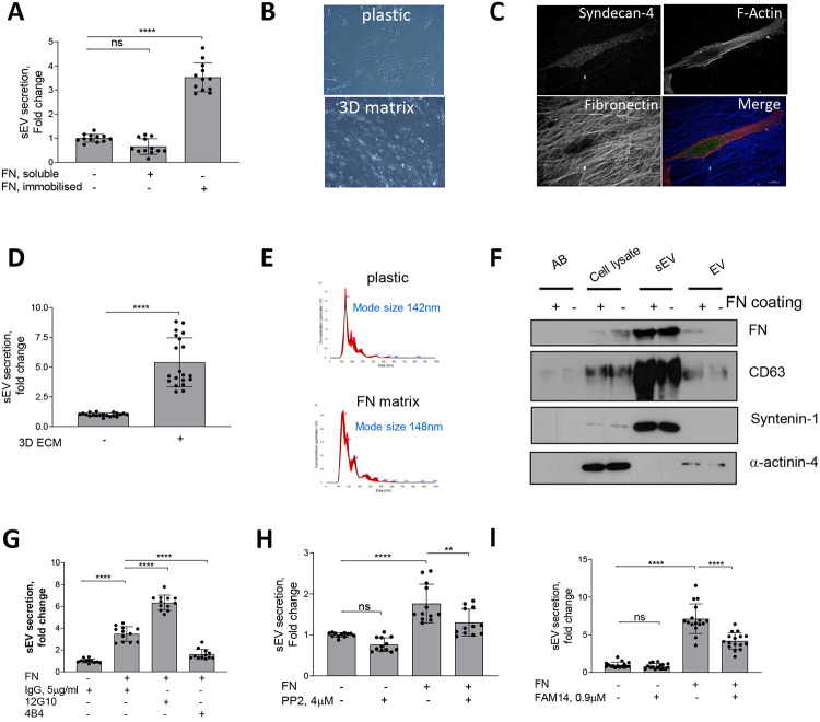
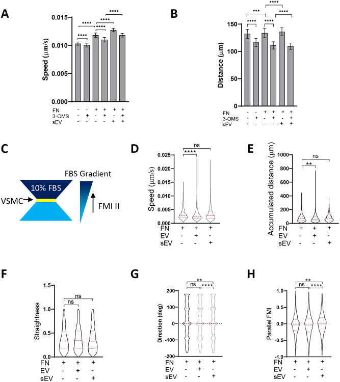
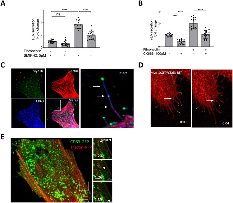
The extracellular matrix (ECM) supports blood vessel architecture and functionality and undergoes active remodelling during vascular repair and atherogenesis. Vascular smooth muscle cells (VSMCs) are essential for vessel repair and, via their secretome, are able to invade from the vessel media into the intima to mediate ECM remodelling. Accumulation of fibronectin (FN) is a hallmark of early vascular repair and atherosclerosis and here we show that FN stimulates VSMCs to secrete small extracellular vesicles (sEVs) by activating the β1 integrin/FAK/Src pathway as well as Arp2/3-dependent branching of the actin cytoskeleton. Spatially, sEV were secreted via filopodia-like cellular protrusions at the leading edge of migrating cells. We found that sEVs are trapped by the ECM and colocalise with FN in symptomatic atherosclerotic plaques . Functionally, ECM-trapped sEVs induced the formation of focal adhesions (FA) with enhanced pulling forces at the cellular periphery. Proteomic and GO pathway analysis revealed that VSMC-derived sEVs display a cell adhesion signature and are specifically enriched with collagen VI. assays identified collagen VI as playing the key role in cell adhesion and invasion. Taken together our data suggests that the accumulation of FN is a key early event in vessel repair acting to promote secretion of collage VI enriched sEVs by VSMCs. These sEVs stimulate migration and invasion by triggering peripheral focal adhesion formation and actomyosin contraction to exert sufficient traction forces to enable VSMC movement within the complex vascular ECM network.
细胞外基质(ECM)支持血管结构和功能,并在血管修复和动脉粥样硬化形成过程中经历活跃的重塑。血管平滑肌细胞(VSMC)对血管修复至关重要,并且通过其分泌组能够从血管中膜侵入内膜以介导ECM重塑。纤连蛋白(FN)的积累是早期血管修复和动脉粥样硬化的标志,在这里我们表明FN通过激活β1整合素/FAK/Src途径以及肌动蛋白细胞骨架的Arp2/3依赖性分支来刺激VSMC分泌小细胞外囊泡(sEV)。在空间上,sEV通过迁移细胞前缘的丝状伪足样细胞突起分泌。我们发现sEV被ECM捕获,并在有症状的动脉粥样硬化斑块中与FN共定位。在功能上,ECM捕获的sEV诱导了粘着斑(FA)的形成,在细胞周边具有增强的拉力。蛋白质组学和GO途径分析表明,VSMC衍生的sEV显示出细胞粘附特征,并特异性富集胶原蛋白VI。实验确定胶原蛋白VI在细胞粘附和侵袭中起关键作用。综上所述,我们的数据表明FN的积累是血管修复中的一个关键早期事件,其作用是促进VSMC分泌富含胶原蛋白VI的sEV。这些sEV通过触发外周粘着斑形成和肌动球蛋白收缩来刺激迁移和侵袭,以施加足够的牵引力,使VSMC能够在复杂的血管ECM网络中移动。