Efromson John, Ferrero Giuliano, Bègue Aurélien, Doman Thomas Jedidiah Jenks, Dugo Clay, Barker Andi, Saliu Veton, Reamey Paul, Kim Kanghyun, Harfouche Mark, Yoder Jeffrey A
Ramona Optics Inc., Durham, NC.
Department of Molecular Biological Sciences, North Carolina State University, Raleigh, NC.
bioRxiv. 2023 Aug 18:2023.08.16.553550. doi: 10.1101/2023.08.16.553550.
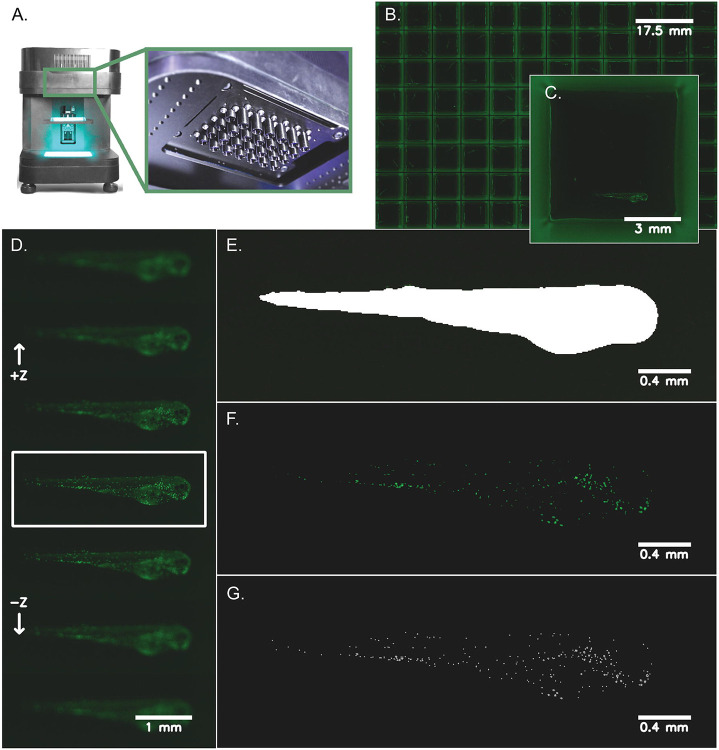
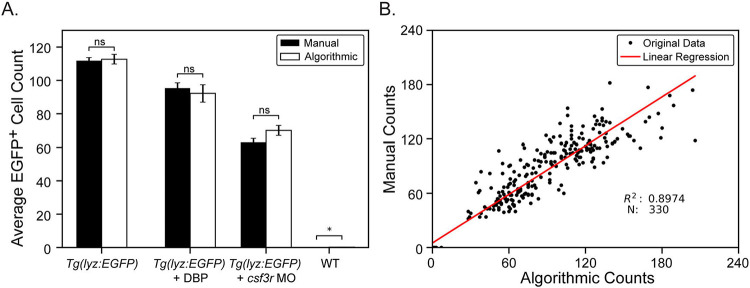
Normal development of the immune system is essential for overall health and disease resistance. Bony fish, such as the zebrafish (), possess all the major immune cell lineages as mammals and can be employed to model human host response to immune challenge. Zebrafish neutrophils, for example, are present in the transparent larvae as early as 48 hours post fertilization and have been examined in numerous infection and immunotoxicology reports. One significant advantage of the zebrafish model is the ability to affordably generate high numbers of individual larvae that can be arrayed in multi-well plates for high throughput genetic and chemical exposure screens. However, traditional workflows for imaging individual larvae have been limited to low-throughput studies using traditional microscopes and manual analyses. Using a newly developed, parallelized microscope, the Multi-Camera Array Microscope (MCAM), we have optimized a rapid, high-resolution algorithmic method to count fluorescently labeled cells in zebrafish larvae . Using transgenic zebrafish larvae, in which neutrophils express EGFP, we captured 18 gigapixels of images across a full 96-well plate, in 75 seconds, and processed the resulting datastream, counting individual fluorescent neutrophils in all individual larvae in 5 minutes. This automation is facilitated by a machine learning segmentation algorithm that defines the most in-focus view of each larva in each well after which pixel intensity thresholding and blob detection are employed to locate and count fluorescent cells. We validated this method by comparing algorithmic neutrophil counts to manual counts in larvae subjected to changes in neutrophil numbers, demonstrating the utility of this approach for high-throughput genetic and chemical screens where a change in neutrophil number is an endpoint metric. Using the MCAM we have been able to, within minutes, acquire both enough data to create an automated algorithm and execute a biological experiment with statistical significance. Finally, we present this open-source software package which allows the user to train and evaluate a custom machine learning segmentation model and use it to localize zebrafish and analyze cell counts within the segmented region of interest. This software can be modified as needed for studies involving other zebrafish cell lineages using different transgenic reporter lines and can also be adapted for studies using other amenable model species.
免疫系统的正常发育对于整体健康和抗病能力至关重要。硬骨鱼,如斑马鱼(),拥有与哺乳动物相同的所有主要免疫细胞谱系,可用于模拟人类宿主对免疫挑战的反应。例如,斑马鱼中性粒细胞在受精后48小时就出现在透明的幼虫中,并且在众多感染和免疫毒理学报告中都有研究。斑马鱼模型的一个显著优点是能够经济地产生大量个体幼虫,这些幼虫可以排列在多孔板中用于高通量遗传和化学暴露筛选。然而,对单个幼虫进行成像的传统工作流程仅限于使用传统显微镜和手动分析的低通量研究。使用新开发的并行显微镜——多相机阵列显微镜(MCAM),我们优化了一种快速、高分辨率的算法方法来计数斑马鱼幼虫中荧光标记的细胞。使用中性粒细胞表达EGFP的转基因斑马鱼幼虫,我们在75秒内跨整个96孔板捕获了180亿像素的图像,并处理了所得数据流,在5分钟内对所有单个幼虫中的单个荧光中性粒细胞进行计数。这种自动化得益于一种机器学习分割算法,该算法定义了每个孔中每个幼虫的最聚焦视图,之后采用像素强度阈值化和斑点检测来定位和计数荧光细胞。我们通过将算法中性粒细胞计数与中性粒细胞数量发生变化的幼虫中的手动计数进行比较,验证了该方法,证明了这种方法在高通量遗传和化学筛选中的实用性,其中中性粒细胞数量的变化是一个终点指标。使用MCAM,我们能够在几分钟内获取足够的数据来创建一个自动化算法,并进行具有统计学意义的生物学实验。最后,我们展示了这个开源软件包,它允许用户训练和评估一个定制的机器学习分割模型,并使用它来定位斑马鱼并分析感兴趣的分割区域内的细胞计数。该软件可以根据需要进行修改,以用于涉及使用不同转基因报告系的其他斑马鱼细胞谱系的研究,也可以适用于使用其他合适模型物种的研究。