School of Medicine, College of Medicine, Kaohsiung Medical University, Kaohsiung 807, Taiwan.
Graduate Institute of Medicine, College of Medicine, Kaohsiung Medical University, Kaohsiung 807, Taiwan.
Theranostics. 2023 Aug 6;13(13):4412-4429. doi: 10.7150/thno.85084. eCollection 2023.
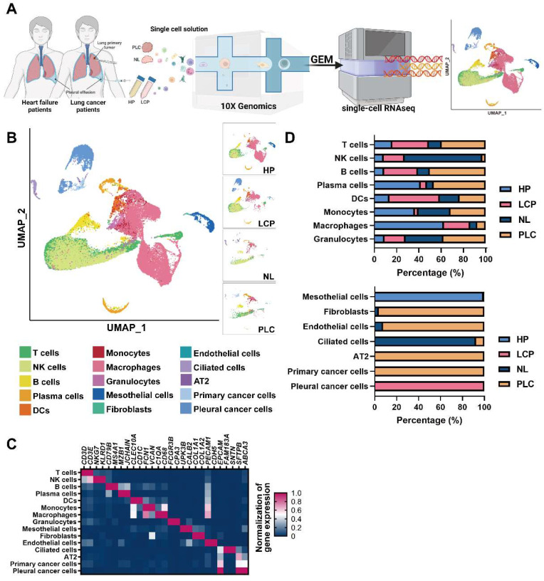
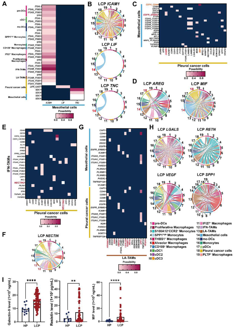
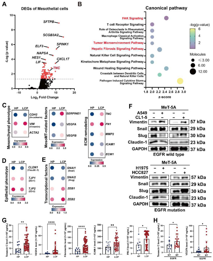
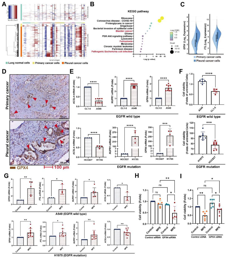
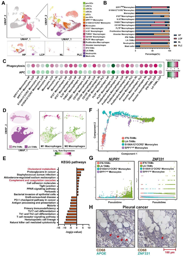
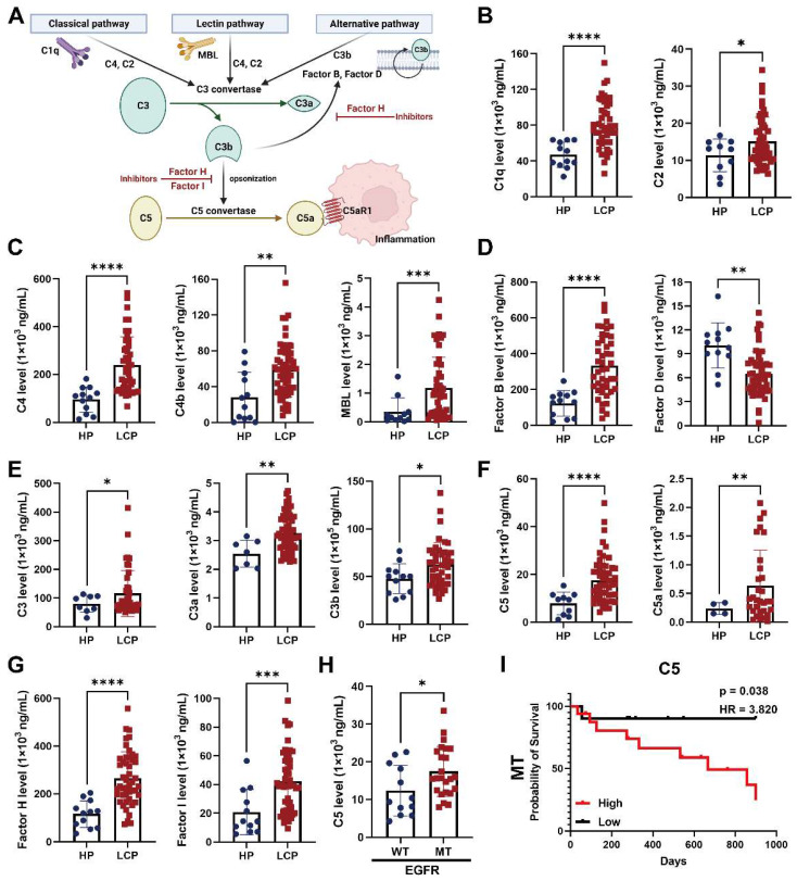
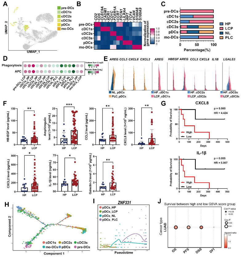
Lung cancer is associated with a high mortality rate and often complicated with malignant pleural effusion (MPE), which has a very poor clinical outcome with a short life expectancy. However, our understanding of cell-specific mechanisms underlying the pathobiology of pleural metastasis remains incomplete. We analyzed single-cell transcriptomes of cells in pleural effusion collected from patients with lung cancer and congestive heart failure (as a control), respectively. Soluble and complement factors were measured using a multiplex cytokine bead assay. The role of ferroptosis was evaluated by small interfering RNA (siRNA) transfection and overexpression. We found that the mesothelial-mesenchymal transition (MesoMT) of the pleural mesothelial cells contributed to pleural metastasis, which was validated by lung cancer/mesothelial cell co-culture experiments. The ferroptosis resistance that protected cancer from death which was secondary to extracellular matrix detachment was critical for pleural metastasis. We found a universal presence of immune-suppressive lipid-associated tumor-associated macrophages (LA-TAMs) with complement cascade alteration in the MPE of the lung cancer patients. Specifically, upregulated complement factors were also found in the MPE, and C5 was associated with poor overall survival in the lung cancer patients with epidermal growth factor receptor mutation. Plasmacytoid dendritic cells (pDCs) exhibited a dysfunctional phenotype and pro-tumorigenic feature in the primary cancer. High expression of the gene set extracted from pDCs was associated with a poor prognosis in the lung cancer patients. Receptor-ligand interaction analysis revealed that the pleural metastatic niche was aggravated by cross-talk between mesothelial cells-cancer cells/immune cells via and . Taken together, our results highlight cell-specific mechanisms involved in the pathobiological development of pleural metastasis in lung cancer. These results provide a large-scale and high-dimensional characterization of the pleural microenvironment and offer a useful resource for the future development of therapeutic drugs in lung cancer.
肺癌死亡率高,常并发恶性胸腔积液(MPE),预期寿命短,临床预后极差。然而,我们对胸腔转移的细胞特异性机制的理解还不完整。我们分别分析了来自肺癌和充血性心力衰竭(作为对照)患者胸腔积液中细胞的单细胞转录组。使用多重细胞因子珠分析测定可溶性和补体因子。通过小干扰 RNA(siRNA)转染和过表达评估铁死亡的作用。我们发现胸膜间皮细胞的间皮-间充质转化(MesoMT)有助于胸腔转移,这在肺癌/间皮细胞共培养实验中得到了验证。铁死亡抵抗对于保护癌症免受细胞外基质脱落后的死亡至关重要,这是胸腔转移所必需的。我们发现,在肺癌患者的胸腔积液中,普遍存在具有补体级联改变的免疫抑制性脂相关肿瘤相关巨噬细胞(LA-TAMs)。具体而言,上调的补体因子也在胸腔积液中发现,C5 与表皮生长因子受体突变的肺癌患者的总生存期不良相关。浆细胞样树突状细胞(pDC)在原发性癌症中表现出功能障碍表型和促肿瘤特征。pDC 中基因集的高表达与肺癌患者的预后不良相关。受体-配体相互作用分析显示,通过间皮细胞-癌细胞/免疫细胞之间的相互作用,胸膜转移灶通过和 加剧。综上所述,我们的研究结果强调了肺癌胸腔转移病理生物学发展中涉及的细胞特异性机制。这些结果为胸腔微环境的大规模、高维特征提供了描述,并为未来肺癌治疗药物的开发提供了有用的资源。