Kolte Vrinda Sunil, Shenoi Ramakrishna S, Dhok Avinash
Department of Oral and Maxillofacial Surgery, VSPM Dental College, Digdoh Hills, Nagpur, Maharashtra, India.
Department of Radiodiagnosis, NKP Salve Institute of Medical Sciences, Digdoh Hills, Nagpur, Maharashtra, India.
Natl J Maxillofac Surg. 2023 May-Aug;14(2):271-276. doi: 10.4103/njms.njms_477_21. Epub 2023 Jul 13.
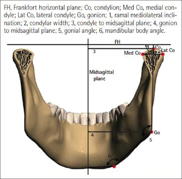
The temporomandibular joint (TMJ) is a unique structure of the body where the mandible, one of the important facial bones, articulates with the temporal part of the skull bone. Obtaining morphometric dimensions for mandibular condyle is important for performing an accurate pre/postoperative assessment, planning temporomandibular and orthognathic surgeries, and applications in forensic sciences in context to the Indian population, which is presently based on dimensions of Caucasian population from available literature. Several investigators noticed the variation in the craniofacial morphology in different ethnic groups and vary according to age and sex. This study aims to provide the normal dimensions of the mandibular condyle in the Indian population, which would be providing racially specific values for diagnosis, treatment planning of surgeries involving condylar processes such as rigid internal fixation of TMJ region, congenital deformities, and customizing TMJ prosthesis concerning these measurements.
To measure the change in dimensions of mandibular condyle according to age and sex using computed topographic scan imaging.
A retrospective analytical cohort study.
Indian adult males and females aged between 20 and 50 years who underwent facial computed tomography (CT) for any reason (e.g., head injury).
Patients with congenital or acquired dentofacial deformities involving TMJ.
By assessing the morphometric dimensions of condyle of mandible using CT scan images.
RESULT/CONCLUSION: Mean condylar dimensions for each age/sex cohort are established; however, no significant change as per age and sex in condylar dimensions in the Indian population is noted.
颞下颌关节(TMJ)是人体独特的结构,其中重要的面骨之一下颌骨与颅骨的颞部相连。获取下颌髁突的形态测量尺寸对于进行准确的术前/术后评估、颞下颌及正颌外科手术规划以及在法医学中针对印度人群的应用非常重要,目前这些应用基于现有文献中白种人的尺寸。一些研究人员注意到不同种族群体的颅面形态存在差异,且会因年龄和性别而有所不同。本研究旨在提供印度人群下颌髁突的正常尺寸,这将为涉及髁突的手术(如颞下颌关节区域的坚固内固定、先天性畸形)的诊断、治疗规划以及根据这些测量定制颞下颌关节假体提供种族特异性值。
使用计算机断层扫描成像测量下颌髁突尺寸随年龄和性别的变化。
一项回顾性分析队列研究。
因任何原因(如头部受伤)接受面部计算机断层扫描(CT)的20至50岁印度成年男性和女性。
患有涉及颞下颌关节的先天性或后天性牙颌面畸形的患者。
通过使用CT扫描图像评估下颌骨髁突的形态测量尺寸。
结果/结论:确定了每个年龄/性别队列的髁突平均尺寸;然而,在印度人群中未发现髁突尺寸随年龄和性别有显著变化。