• 文献检索
  • 文档翻译
  • 深度研究
  • 学术资讯
  • Suppr Zotero 插件Zotero 插件
  • 邀请有礼
  • 套餐&价格
  • 历史记录
应用&插件
Suppr Zotero 插件Zotero 插件浏览器插件Mac 客户端Windows 客户端微信小程序
定价
高级版会员购买积分包购买API积分包
服务
文献检索文档翻译深度研究API 文档MCP 服务
关于我们
关于 Suppr公司介绍联系我们用户协议隐私条款
关注我们

Suppr 超能文献

核心技术专利:CN118964589B侵权必究
粤ICP备2023148730 号-1Suppr @ 2026

文献检索

告别复杂PubMed语法,用中文像聊天一样搜索,搜遍4000万医学文献。AI智能推荐,让科研检索更轻松。

立即免费搜索

文件翻译

保留排版,准确专业,支持PDF/Word/PPT等文件格式,支持 12+语言互译。

免费翻译文档

深度研究

AI帮你快速写综述,25分钟生成高质量综述,智能提取关键信息,辅助科研写作。

立即免费体验

DNA 和组蛋白会损害葡萄球菌凝固酶形成的纤维蛋白的机械稳定性和溶酶体易感性。

DNA and histones impair the mechanical stability and lytic susceptibility of fibrin formed by staphylocoagulase.

机构信息

Institute of Biochemistry and Molecular Biology, Department of Biochemistry, Semmelweis University, Budapest, Hungary.

Plasma Chemistry Research Group, Institute of Materials and Environmental Chemistry, Research Centre for Natural Sciences, Budapest, Hungary.

出版信息

Front Immunol. 2023 Aug 17;14:1233128. doi: 10.3389/fimmu.2023.1233128. eCollection 2023.

DOI:10.3389/fimmu.2023.1233128
PMID:37662916
原文链接:https://pmc.ncbi.nlm.nih.gov/articles/PMC10470048/
Abstract

BACKGROUND

Staphylocoagulase (SCG) is a virulence factor of , one of the most lethal pathogens of our times. The complex of SCG with prothrombin (SCG/ProT) can clot fibrinogen, and SCG/ProT-induced fibrin and plasma clots have been described to show decreased mechanical and lytic resistance, which may contribute to septic emboli from infected cardiac vegetations. At infection sites, neutrophils can release DNA and histones, as parts of neutrophil extracellular traps (NETs), which in turn favor thrombosis, inhibit fibrinolysis and strengthen clot structure.

OBJECTIVES

To characterize the combined effects of major NET-components (DNA, histone H1 and H3) on SCG/ProT-induced clot structure, mechanical and lytic stability.

METHODS

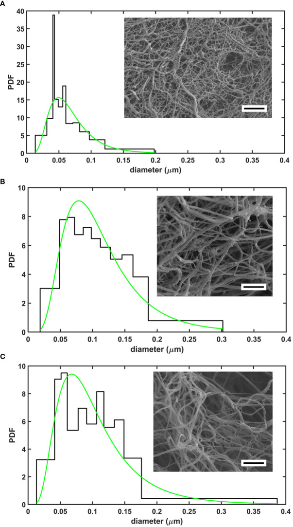
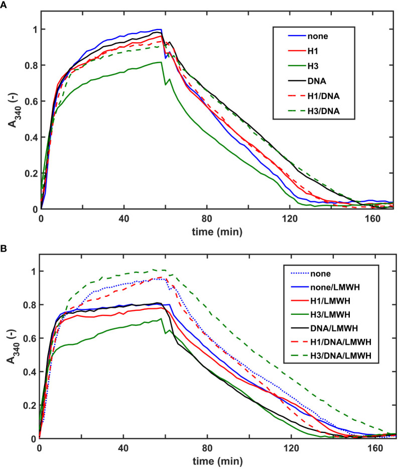
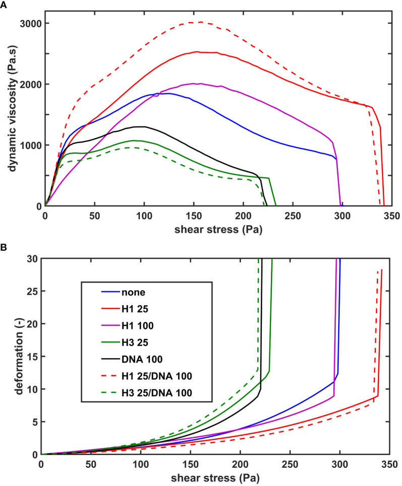
Recombinant SCG was used to clot purified fibrinogen and plasma. The kinetics of formation and lysis of fibrin and plasma clots containing H1 or core histones+/-DNA were followed by turbidimetry. Fibrin structure and mechanical stability were characterized with scanning electron microscopy, pressure-driven permeation, and oscillation rheometry.

RESULTS

Histones and DNA favored the formation of thicker fibrin fibers and a more heterogeneous clot structure including high porosity with H1 histone, whereas low porosity with core histones and DNA. As opposed to previous observations with thrombin-induced clots, SCG/ProT-induced fibrin was not mechanically stabilized by histones. Similarly to thrombin-induced clots, the DNA-histone complexes prolonged fibrinolysis with tissue-type plasminogen activator (up to 2-fold). The anti-fibrinolytic effect of the DNA and DNA-H3 complex was observed in plasma clots too. Heparin (low molecular weight) accelerated the lysis of SCG/ProT-clots from plasma, even if DNA and histones were also present.

CONCLUSIONS

In the interplay of NETs and fibrin formed by SCG, DNA and histones promote structural heterogeneity in the clots, and fail to stabilize them against mechanical stress. The DNA-histone complexes render the SCG-fibrin more resistant to lysis and thereby less prone to embolization.

摘要

背景

凝固酶(SCG)是葡萄球菌的一种毒力因子,也是当今最致命的病原体之一。SCG 与凝血酶原(SCG/ProT)的复合物可以使纤维蛋白原凝结,并且已经描述了 SCG/ProT 诱导的纤维蛋白和血浆凝块显示出机械和溶解稳定性降低,这可能导致感染性心内膜炎的心脏赘生物中的感染性栓子。在感染部位,中性粒细胞可以释放 DNA 和组蛋白,作为中性粒细胞细胞外陷阱(NETs)的一部分,这反过来又有利于血栓形成、抑制纤维蛋白溶解并增强凝块结构。

目的

描述主要 NET 成分(DNA、组蛋白 H1 和 H3)对 SCG/ProT 诱导的凝块结构、机械和溶解稳定性的综合影响。

方法

使用重组 SCG 使纯化的纤维蛋白原和血浆凝结。通过浊度法跟踪 H1 组蛋白或核心组蛋白+/-DNA 存在时纤维蛋白和血浆凝块形成和溶解的动力学。通过扫描电子显微镜、压力驱动渗透和振荡流变学来表征纤维蛋白结构和机械稳定性。

结果

组蛋白和 DNA 有利于形成更厚的纤维蛋白纤维和更不均匀的凝块结构,包括 H1 组蛋白的高孔隙率,而核心组蛋白和 DNA 的孔隙率低。与以前用凝血酶诱导的凝块观察到的情况相反,组蛋白没有使 SCG/ProT 诱导的纤维蛋白机械稳定。与凝血酶诱导的凝块一样,DNA-组蛋白复合物延长了组织型纤溶酶原激活剂(最多 2 倍)诱导的纤维蛋白溶解。DNA 和 DNA-H3 复合物也在血浆凝块中观察到抗纤维蛋白溶解作用。肝素(低分子量)加速了来自血浆的 SCG/ProT 凝块的溶解,即使存在 DNA 和组蛋白也是如此。

结论

在 NETs 和 SCG 形成的纤维蛋白的相互作用中,DNA 和组蛋白促进凝块结构的异质性,并且不能使它们抵抗机械应力。DNA-组蛋白复合物使 SCG 纤维蛋白更能抵抗溶解,从而不易栓塞。

https://cdn.ncbi.nlm.nih.gov/pmc/blobs/995d/10470048/7e16f94185cb/fimmu-14-1233128-g004.jpg
https://cdn.ncbi.nlm.nih.gov/pmc/blobs/995d/10470048/fe01902b7d4a/fimmu-14-1233128-g001.jpg
https://cdn.ncbi.nlm.nih.gov/pmc/blobs/995d/10470048/de2eefc2dbd9/fimmu-14-1233128-g002.jpg
https://cdn.ncbi.nlm.nih.gov/pmc/blobs/995d/10470048/241b0504a1c9/fimmu-14-1233128-g003.jpg
https://cdn.ncbi.nlm.nih.gov/pmc/blobs/995d/10470048/7e16f94185cb/fimmu-14-1233128-g004.jpg

相似文献

1
DNA and histones impair the mechanical stability and lytic susceptibility of fibrin formed by staphylocoagulase.DNA 和组蛋白会损害葡萄球菌凝固酶形成的纤维蛋白的机械稳定性和溶酶体易感性。
Front Immunol. 2023 Aug 17;14:1233128. doi: 10.3389/fimmu.2023.1233128. eCollection 2023.
2
Structure, Mechanical, and Lytic Stability of Fibrin and Plasma Coagulum Generated by Staphylocoagulase From .由金黄色葡萄球菌凝固酶生成的纤维蛋白和血浆凝块的结构、力学和溶解释放稳定性。
Front Immunol. 2019 Dec 20;10:2967. doi: 10.3389/fimmu.2019.02967. eCollection 2019.
3
Size- and charge-dependent modulation of the lytic susceptibility and mechanical stability of fibrin-histone clots by heparin and polyphosphate variants.肝素和多聚磷酸盐变体对纤维蛋白-组蛋白凝块的溶解释放敏感性和机械稳定性的尺寸和电荷依赖性调节。
J Thromb Haemost. 2021 May;19(5):1307-1318. doi: 10.1111/jth.15258. Epub 2021 Feb 20.
4
DNA, histones and neutrophil extracellular traps exert anti-fibrinolytic effects in a plasma environment.DNA、组蛋白和中性粒细胞胞外诱捕网在血浆环境中发挥抗纤溶作用。
Thromb Haemost. 2015 Jun;113(6):1289-98. doi: 10.1160/TH14-08-0669. Epub 2015 Mar 19.
5
Mechanical stability and fibrinolytic resistance of clots containing fibrin, DNA, and histones.含有纤维蛋白、DNA 和组蛋白的血栓的机械稳定性和抗纤维蛋白溶解能力。
J Biol Chem. 2013 Mar 8;288(10):6946-56. doi: 10.1074/jbc.M112.404301. Epub 2013 Jan 4.
6
Biorelevant polyanions stabilize fibrin against mechanical and proteolytic decomposition: Effects of polymer size and electric charge.具有生物相关性的聚阴离子可稳定纤维蛋白,抵抗机械和蛋白水解分解:聚合物大小和电荷的影响。
J Mech Behav Biomed Mater. 2020 Feb;102:103459. doi: 10.1016/j.jmbbm.2019.103459. Epub 2019 Sep 28.
7
Polyphosphate nanoparticles enhance the fibrin stabilization by histones more efficiently than linear polyphosphates.多聚磷酸盐纳米颗粒比线性多聚磷酸盐更有效地增强组蛋白稳定纤维蛋白的能力。
PLoS One. 2022 Apr 25;17(4):e0266782. doi: 10.1371/journal.pone.0266782. eCollection 2022.
8
Canine Neutrophil Extracellular Traps Enhance Clot Formation and Delay Lysis.犬中性粒细胞胞外诱捕网增强血栓形成并延迟溶解。
Vet Pathol. 2018 Jan;55(1):116-123. doi: 10.1177/0300985817699860. Epub 2017 Mar 27.
9
Cell-Free DNA Modulates Clot Structure and Impairs Fibrinolysis in Sepsis.无细胞游离 DNA 调节脓毒症中的血栓结构并损害纤维蛋白溶解。
Arterioscler Thromb Vasc Biol. 2015 Dec;35(12):2544-53. doi: 10.1161/ATVBAHA.115.306035. Epub 2015 Oct 22.
10
Citrullinated fibrinogen forms densely packed clots with decreased permeability.瓜氨酸化纤维蛋白原形成致密的血栓,其通透性降低。
J Thromb Haemost. 2022 Dec;20(12):2862-2872. doi: 10.1111/jth.15875. Epub 2022 Sep 27.

引用本文的文献

1
Recent Advances in Pathogenesis and Anticoagulation Treatment of Sepsis-Induced Coagulopathy.脓毒症诱导的凝血病发病机制及抗凝治疗的最新进展
J Inflamm Res. 2025 Jan 18;18:737-750. doi: 10.2147/JIR.S495223. eCollection 2025.
2
Deconstructing fibrin(ogen) structure.解析纤维蛋白(原)结构。
J Thromb Haemost. 2025 Feb;23(2):368-380. doi: 10.1016/j.jtha.2024.10.024. Epub 2024 Nov 12.

本文引用的文献

1
Neutrophils Protect Against Endocarditis Progression Independent of Extracellular Trap Release.中性粒细胞通过不依赖于细胞外陷阱释放来保护对抗心内膜炎进展。
Arterioscler Thromb Vasc Biol. 2023 Feb;43(2):267-285. doi: 10.1161/ATVBAHA.122.317800. Epub 2022 Dec 1.
2
Infective Endocarditis Remains a Deadly Disease-It's Bad News, Especially When and Are Involved: A Call to Action.感染性心内膜炎仍然是一种致命疾病——这是个坏消息,尤其是当[此处原文缺失两个关键信息]涉及其中时:呼吁采取行动。
J Am Heart Assoc. 2022 Aug 16;11(16):e026788. doi: 10.1161/JAHA.122.026788. Epub 2022 Aug 10.
3
Temporal Changes, Patient Characteristics, and Mortality, According to Microbiological Cause of Infective Endocarditis: A Nationwide Study.
根据感染性心内膜炎的微生物学病因的时间变化、患者特征和死亡率:一项全国性研究。
J Am Heart Assoc. 2022 Aug 16;11(16):e025801. doi: 10.1161/JAHA.122.025801. Epub 2022 Aug 10.
4
Inhibition of NETosis for treatment purposes: friend or foe?抑制 NETosis 用于治疗:是敌是友?
Mol Cell Biochem. 2022 Mar;477(3):673-688. doi: 10.1007/s11010-021-04315-x. Epub 2022 Jan 7.
5
Use of direct oral anticoagulants in ICU patients. Part II - Clinical evidence.ICU 患者中直接口服抗凝剂的应用。第二部分 - 临床证据。
Anaesthesiol Intensive Ther. 2021;53(5):440-449. doi: 10.5114/ait.2021.110608.
6
Quantitative analysis of clot density, fibrin fiber radius, and protofibril packing in acute phase myocardial infarction.对急性期心肌梗死中血栓密度、纤维蛋白纤维半径和原纤维堆积的定量分析。
Thromb Res. 2021 Sep;205:110-119. doi: 10.1016/j.thromres.2021.06.024. Epub 2021 Jul 8.
7
Anti-biofilm Approach in Infective Endocarditis Exposes New Treatment Strategies for Improved Outcome.感染性心内膜炎中的抗生物膜方法揭示了改善预后的新治疗策略。
Front Cell Dev Biol. 2021 Jun 18;9:643335. doi: 10.3389/fcell.2021.643335. eCollection 2021.
8
induces neutrophil extracellular traps (NETs) and neutralizes their bactericidal potential.诱导中性粒细胞胞外诱捕网(NETs)并中和其杀菌潜力。
Comput Struct Biotechnol J. 2021 Jun 6;19:3451-3457. doi: 10.1016/j.csbj.2021.06.012. eCollection 2021.
9
Current Challenges in the Management of Infective Endocarditis.感染性心内膜炎管理中的当前挑战
Front Med (Lausanne). 2021 Feb 22;8:641243. doi: 10.3389/fmed.2021.641243. eCollection 2021.
10
Size- and charge-dependent modulation of the lytic susceptibility and mechanical stability of fibrin-histone clots by heparin and polyphosphate variants.肝素和多聚磷酸盐变体对纤维蛋白-组蛋白凝块的溶解释放敏感性和机械稳定性的尺寸和电荷依赖性调节。
J Thromb Haemost. 2021 May;19(5):1307-1318. doi: 10.1111/jth.15258. Epub 2021 Feb 20.