Program in Cellular and Molecular Medicine, Boston Children's Hospital, Boston, Massachusetts, USA.
Department of Pediatrics, Harvard Medical School, Boston, Massachusetts, USA.
J Thromb Haemost. 2022 Dec;20(12):2862-2872. doi: 10.1111/jth.15875. Epub 2022 Sep 27.
Fibrin, the main scaffold of thrombi, is susceptible to citrullination by PAD (peptidyl arginine deiminase) 4, secreted from neutrophils during the formation of neutrophil extracellular traps. Citrullinated fibrinogen (citFg) has been detected in human plasma as well as in murine venous thrombi, and it decreases the lysability and mechanical resistance of fibrin clots.
To investigate the effect of fibrinogen citrullination on the structure of fibrin clots.
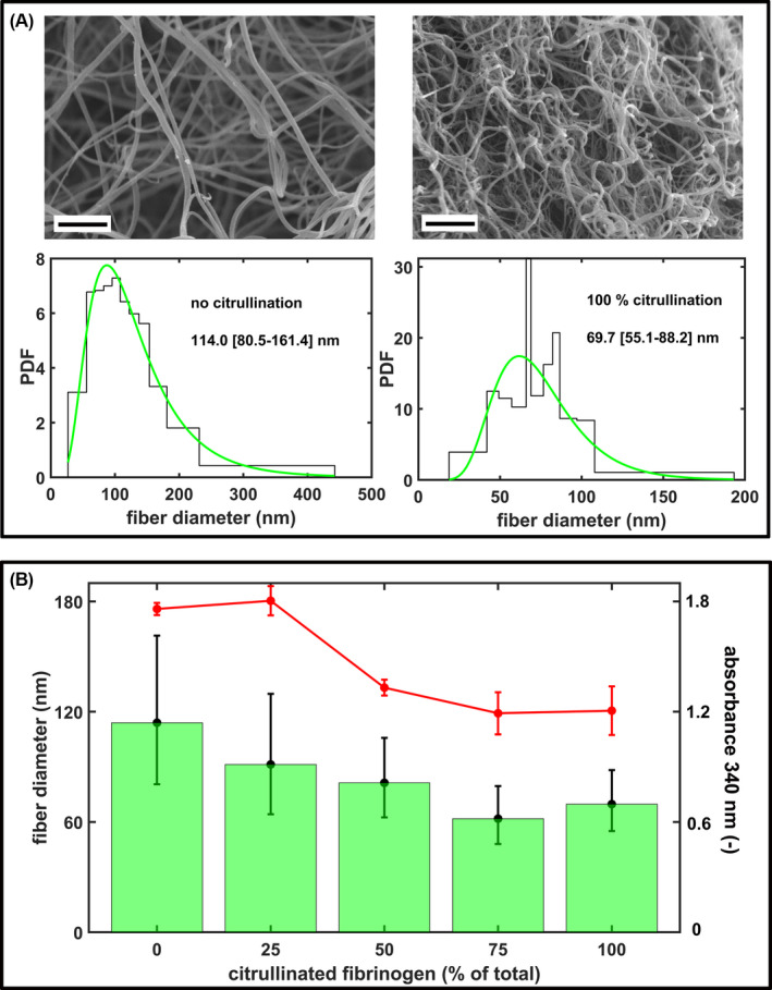
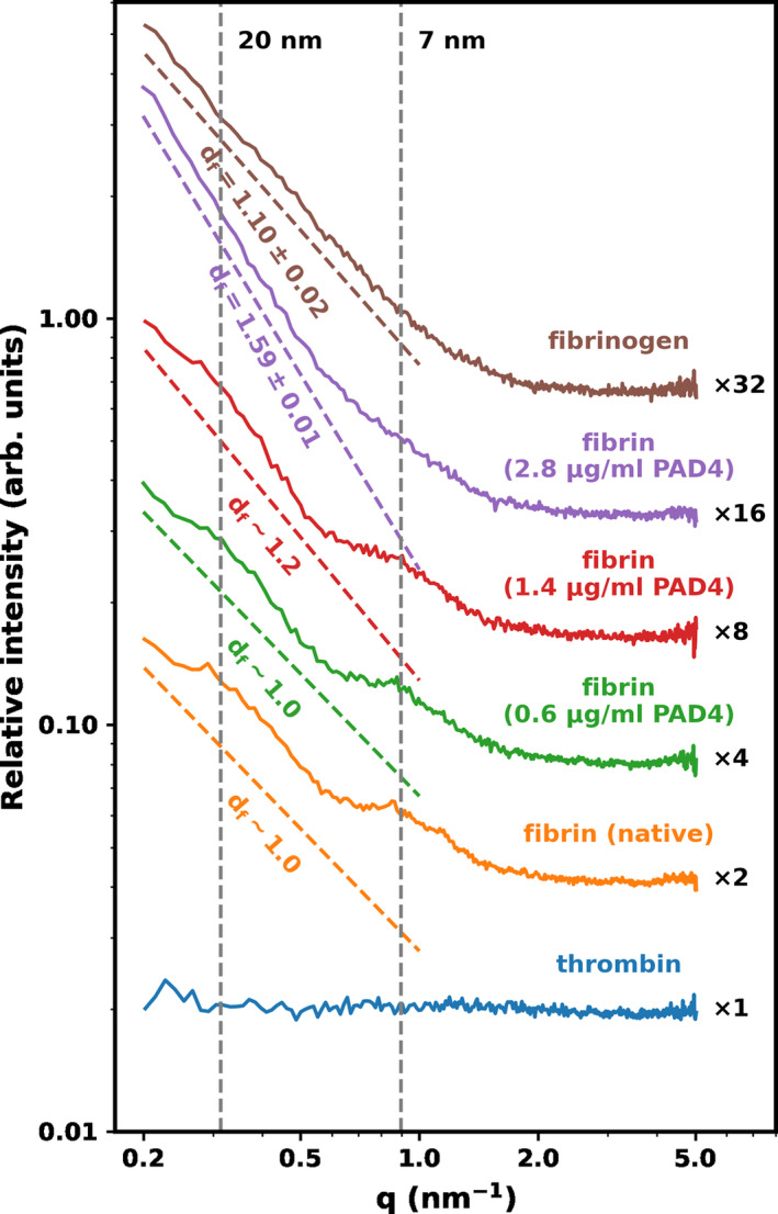
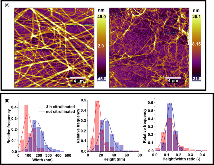
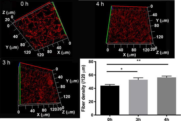
Fibrinogen was citrullinated with PAD4 and clotted with thrombin. Scanning electron microscopy (SEM) and atomic force microscopy (AFM) were used to measure fiber thickness, fiber height/width ratio, and fiber persistence length in clots containing citFg. Fiber density was measured with laser scanning microscopy (LSM) and permeability measurements were carried out to estimate the porosity of the clots. The intra-fiber structure of fibrin was analyzed with small-angle X-ray scattering (SAXS).
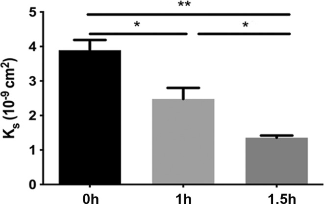
SEM images revealed a decrease in the median fiber diameter that correlated with the fraction of citFg in the clot, while the fiber width/length ratio remained unchanged according to AFM. With SAXS we observed that citrullination resulted in the formation of denser clots in line with increased fiber density shown by LSM. The permeability constant of citrullinated fibrin decreased more than 3-fold indicating significantly decreased porosity. SAXS also showed largely preserved periodicity in the longitudinal assembly of fibrin monomers.
The current observations of thin fibers combined with dense packing and low porosity in the presence of citFg can provide a structural framework for the mechanical fragility and lytic resistance of citrullinated fibrin.
纤维蛋白是血栓的主要支架,易被中性粒细胞在形成中性粒细胞胞外诱捕网时分泌的 PAD(肽基精氨酸脱亚氨酶)4 瓜氨酸化。瓜氨酸化纤维蛋白原(citFg)已在人血浆和鼠静脉血栓中检测到,它降低了纤维蛋白凝块的可裂解性和机械阻力。
研究纤维蛋白原瓜氨酸化对纤维蛋白凝块结构的影响。
用 PAD4 对纤维蛋白原进行瓜氨酸化,然后用凝血酶进行凝结。扫描电子显微镜(SEM)和原子力显微镜(AFM)用于测量含有 citFg 的凝块中纤维的厚度、纤维高度/宽度比和纤维持久长度。激光扫描显微镜(LSM)用于测量纤维密度,渗透率测量用于估计凝块的孔隙率。用小角 X 射线散射(SAXS)分析纤维内的纤维蛋白结构。
SEM 图像显示,纤维的中值直径随着凝块中 citFg 的比例降低而降低,而纤维的宽度/长度比根据 AFM 保持不变。用 SAXS 观察到,瓜氨酸化导致凝块更致密,这与 LSM 所示的纤维密度增加一致。瓜氨酸化纤维的渗透率常数降低了 3 倍以上,表明孔隙率显著降低。SAXS 还显示纤维单体的纵向组装中保留了很大的周期性。
目前观察到的纤维较细,结合存在 citFg 时的致密堆积和低孔隙率,可以为瓜氨酸化纤维的机械脆弱性和裂解抗性提供结构框架。