Ball Jayson B, McNulty Connor J, Green-Fulgham Suzanne M, Dragavon Joseph M, Correia Rocha Igor R, Finch Maggie R, Prévost Emily D, Siddique Imaad I, Woodall Brodie J, Watkins Linda R, Baratta Michael V, Root David H
Department of Psychology and Neuroscience, Center for Neuroscience, University of Colorado Boulder, Boulder, CO, United States.
Advanced Light Microscopy Core, Biofrontiers Institute, University of Colorado Boulder, Boulder, CO, United States.
Front Mol Neurosci. 2023 Aug 17;16:1225847. doi: 10.3389/fnmol.2023.1225847. eCollection 2023.
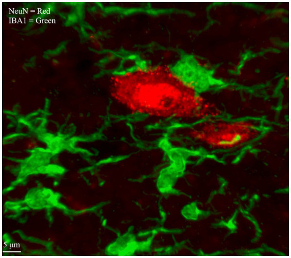
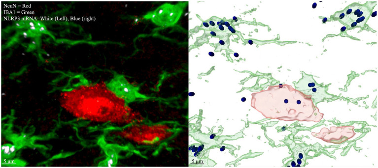
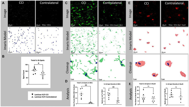
A challenge for central nervous system (CNS) tissue analysis in neuroscience research has been the difficulty to codetect and colocalize gene and protein expression in the same tissue. Given the importance of identifying gene expression relative to proteins of interest, for example, cell-type specific markers, we aimed to develop a protocol to optimize their codetection. RNAscope fluorescent hybridization (FISH) combined with immunohistochemistry (IHC) in fixed (CNS) tissue sections allows for reliable quantification of gene transcripts of interest within IHC-labeled cells. This paper describes a new method for simultaneous visualization of FISH and IHC in thicker (14-μm), fixed tissue samples, using spinal cord sections. This method's effectiveness is shown by the cell-type-specific quantification of two genes, namely the proinflammatory cytokine interleukin-1beta (IL-1b) and the inflammasome NLR family pyrin domain containing 3 (NLRP3). These genes are challenging to measure accurately using immunohistochemistry (IHC) due to the nonspecificity of available antibodies and the hard-to-distinguish, dot-like visualizations of the labeled proteins within the tissue. These measurements were carried out in spinal cord sections after unilateral chronic constriction injury of the sciatic nerve to induce neuroinflammation in the spinal cord. RNAscope is used to label transcripts of genes of interest and IHC is used to label cell-type specific antigens (IBA1 for microglia, NeuN for neurons). This combination allowed for labeled RNA transcripts to be quantified within cell-type specific boundaries using confocal microscopy and standard image analysis methods. This method makes it easy to answer empirical questions that are intractable with standard IHC or hybridization alone. The method, which has been optimized for spinal cord tissue and to minimize tissue preparation time and costs, is described in detail from tissue collection to image analysis. Further, the relative expression changes in inflammatory genes NLRP3 and IL-1b in spinal cord microglia vs. neurons of somatotopically relevant laminae are described for the first time.
神经科学研究中,中枢神经系统(CNS)组织分析面临的一个挑战是难以在同一组织中同时检测和共定位基因与蛋白质表达。鉴于识别相对于感兴趣蛋白质(例如细胞类型特异性标志物)的基因表达的重要性,我们旨在开发一种方案来优化它们的同时检测。固定的(CNS)组织切片中,RNAscope荧光原位杂交(FISH)与免疫组织化学(IHC)相结合,能够在免疫组织化学标记的细胞内可靠地定量感兴趣的基因转录本。本文描述了一种在较厚(14μm)的固定组织样本(使用脊髓切片)中同时可视化FISH和IHC的新方法。通过对两个基因进行细胞类型特异性定量,即促炎细胞因子白细胞介素-1β(IL-1β)和含吡啉结构域的NLR家族成员3(NLRP3),证明了该方法的有效性。由于现有抗体的非特异性以及组织内标记蛋白难以区分的点状可视化,使用免疫组织化学(IHC)准确测量这些基因具有挑战性。这些测量是在坐骨神经单侧慢性压迫损伤后进行的脊髓切片中进行的,以诱导脊髓中的神经炎症。RNAscope用于标记感兴趣基因的转录本,IHC用于标记细胞类型特异性抗原(小胶质细胞用IBA1,神经元用NeuN)。这种组合使得可以使用共聚焦显微镜和标准图像分析方法在细胞类型特异性边界内对标记的RNA转录本进行定量。该方法使得回答仅用标准IHC或杂交难以解决的实证问题变得容易。本文详细描述了该方法,该方法已针对脊髓组织进行了优化,以尽量减少组织制备时间和成本,从组织采集到图像分析。此外,首次描述了脊髓小胶质细胞与躯体感觉相关板层神经元中炎症基因NLRP3和IL-1β的相对表达变化。