Hazama F, Kataoka H, Yamada E, Kayembe K, Hashimoto N, Kojima M, Kim C
Am J Pathol. 1986 Sep;124(3):399-404.
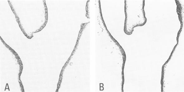
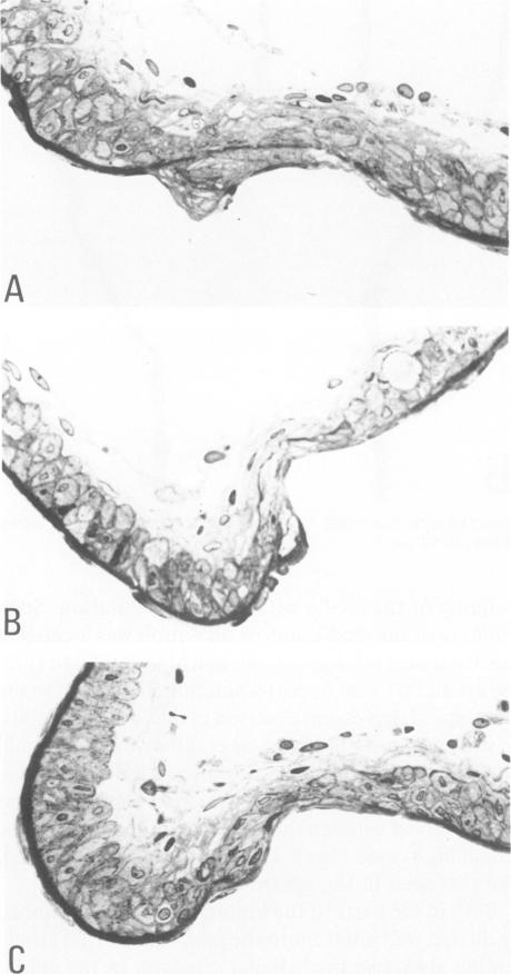
The changes of the anterior cerebral artery/olfactory artery junction, one of the favorite sites of aneurysm formation, in rats treated with unilateral ligation of the common carotid artery and renal hypertension were investigated by light microscopy. The initial changes of aneurysm occurred not at the apex itself, but on the distal side of the major branch adjacent to the apex, at the intimal pad and the neighboring distal portion. Here the internal elastic lamina showed various degenerative changes and disappearance. The neighboring distal portion adjacent to the intimal pad showed a shallow depression associated with a thinning of the media due to a decrease of medial smooth muscle cells in number even in some control animals. Such degenerative changes of the internal elastic lamina and medial smooth muscle cells caused by hemodynamic stress due to branching structure, including intimal pads, augmented by the experimental treatment, are supposed to be the basis for aneurysm formation.
通过光学显微镜研究了单侧颈总动脉结扎和肾性高血压处理的大鼠中,作为动脉瘤形成常见部位之一的大脑前动脉/嗅动脉交界处的变化。动脉瘤的初始变化并非发生在顶端本身,而是在与顶端相邻的主要分支的远端侧,在内膜垫及其相邻的远端部分。此处内弹性膜呈现出各种退行性变化并消失。即使在一些对照动物中,与内膜垫相邻的远端部分也显示出浅凹陷,伴有中膜变薄,这是由于中膜平滑肌细胞数量减少所致。由包括内膜垫在内的分支结构引起的血流动力学应力导致的内弹性膜和中膜平滑肌细胞的这种退行性变化,经实验处理后进一步加剧,被认为是动脉瘤形成的基础。