• 文献检索
  • 文档翻译
  • 深度研究
  • 学术资讯
  • Suppr Zotero 插件Zotero 插件
  • 邀请有礼
  • 套餐&价格
  • 历史记录
应用&插件
Suppr Zotero 插件Zotero 插件浏览器插件Mac 客户端Windows 客户端微信小程序
定价
高级版会员购买积分包购买API积分包
服务
文献检索文档翻译深度研究API 文档MCP 服务
关于我们
关于 Suppr公司介绍联系我们用户协议隐私条款
关注我们

Suppr 超能文献

核心技术专利:CN118964589B侵权必究
粤ICP备2023148730 号-1Suppr @ 2026

文献检索

告别复杂PubMed语法,用中文像聊天一样搜索,搜遍4000万医学文献。AI智能推荐,让科研检索更轻松。

立即免费搜索

文件翻译

保留排版,准确专业,支持PDF/Word/PPT等文件格式,支持 12+语言互译。

免费翻译文档

深度研究

AI帮你快速写综述,25分钟生成高质量综述,智能提取关键信息,辅助科研写作。

立即免费体验

分水岭脑梗死的诊断与治疗:一篇叙述性综述。

Diagnosis and treatment of Watershed strokes: a narrative review.

机构信息

University of Medicine and Pharmacy, Craiova, Romania.

Department of Neurology, Emergency County Hospital, Targu-Jiu, Romania.

出版信息

J Med Life. 2023 Jun;16(6):842-850. doi: 10.25122/jml-2023-0127.

DOI:10.25122/jml-2023-0127
PMID:37675172
原文链接:https://pmc.ncbi.nlm.nih.gov/articles/PMC10478671/
Abstract

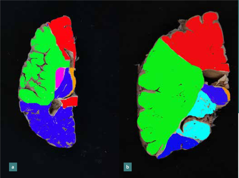
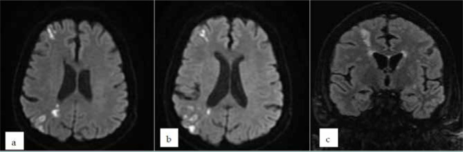
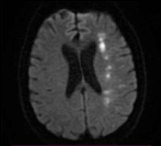
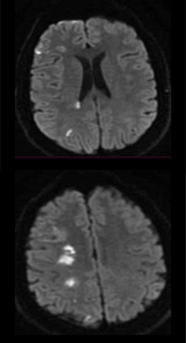
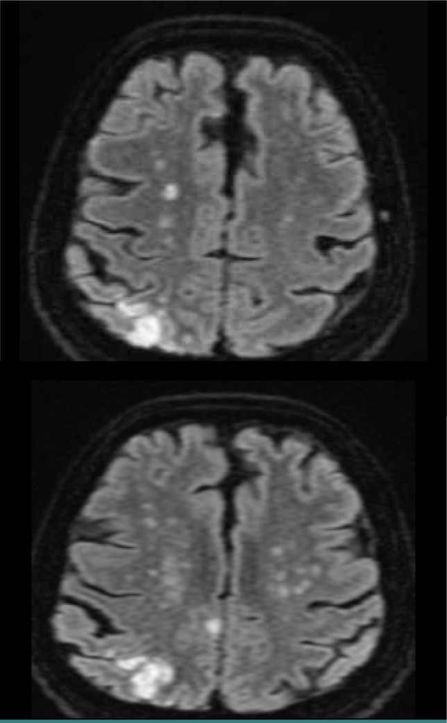
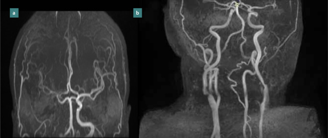
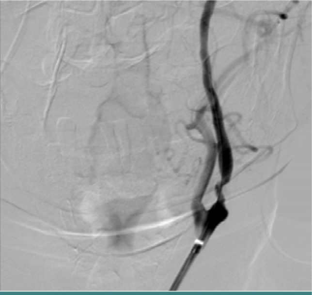
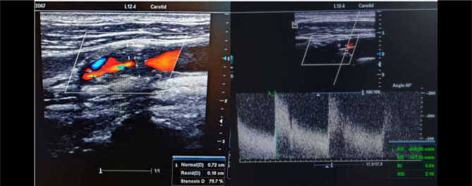
Watershed strokes have been described previously as ischemic strokes located in vulnerable border zones between brain tissue supplied by the anterior, posterior, and middle cerebral arteries in the distal junction between two non-anastomotic arterial territories. Ischemic strokes in border zones are well-recognized entities and well-described in terms of imaging features, but the pathophysiological mechanism of brain injury production is not fully defined. Border zone ischemia is caused by cerebral hypoperfusion through decreased cerebral blood flow and arterial embolism in unstable atheroma plaque. It is often difficult to say which mechanisms are fully responsible for producing cerebral ischemic lesions. This review aimed to highlight the imaging aspect of watershed strokes and to correlate the clinical characteristics of this type of stroke with the diagnostic algorithm for optimal therapeutic management. Neurologists should promptly recognize this type of stroke and investigate its etiology in the shortest possible time.

摘要

分水岭梗死此前被描述为位于大脑前、后、中动脉供血的脑组织之间的交界区,即两个非吻合动脉区域的远端交界处的缺血性脑卒中。交界区的缺血性脑卒中是一种公认的实体,在影像学特征方面有很好的描述,但脑损伤产生的病理生理机制尚未完全确定。交界区的缺血是由于脑灌注减少导致的,其原因包括脑血流量减少和不稳定粥样斑块中的动脉栓塞。通常很难确定哪些机制完全负责产生脑缺血性病变。本综述旨在强调分水岭梗死的影像学表现,并将这种类型的脑卒中的临床特征与最佳治疗管理的诊断算法相关联。神经科医生应该迅速识别这种类型的脑卒中,并在最短的时间内调查其病因。

https://cdn.ncbi.nlm.nih.gov/pmc/blobs/2333/10478671/a8ad6f5ddc0e/JMedLife-16-842-g009.jpg
https://cdn.ncbi.nlm.nih.gov/pmc/blobs/2333/10478671/6895ac68a3be/JMedLife-16-842-g001.jpg
https://cdn.ncbi.nlm.nih.gov/pmc/blobs/2333/10478671/de5ea96351d4/JMedLife-16-842-g002.jpg
https://cdn.ncbi.nlm.nih.gov/pmc/blobs/2333/10478671/a7c5a0376b7e/JMedLife-16-842-g003.jpg
https://cdn.ncbi.nlm.nih.gov/pmc/blobs/2333/10478671/51324d904ff0/JMedLife-16-842-g004.jpg
https://cdn.ncbi.nlm.nih.gov/pmc/blobs/2333/10478671/12c1e647b874/JMedLife-16-842-g005.jpg
https://cdn.ncbi.nlm.nih.gov/pmc/blobs/2333/10478671/f4fd5c75a7e7/JMedLife-16-842-g006.jpg
https://cdn.ncbi.nlm.nih.gov/pmc/blobs/2333/10478671/a22b28a3834d/JMedLife-16-842-g007.jpg
https://cdn.ncbi.nlm.nih.gov/pmc/blobs/2333/10478671/e0a7b094606d/JMedLife-16-842-g008.jpg
https://cdn.ncbi.nlm.nih.gov/pmc/blobs/2333/10478671/a8ad6f5ddc0e/JMedLife-16-842-g009.jpg

相似文献

1
Diagnosis and treatment of Watershed strokes: a narrative review.分水岭脑梗死的诊断与治疗:一篇叙述性综述。
J Med Life. 2023 Jun;16(6):842-850. doi: 10.25122/jml-2023-0127.
2
Patterns of Acute Ischemic Strokes After Carotid Endarterectomy and Therapeutic Implications.颈动脉内膜切除术后急性缺血性卒中的模式及治疗意义
Vasc Endovascular Surg. 2017 Oct;51(7):485-490. doi: 10.1177/1538574417723482. Epub 2017 Aug 28.
3
The relationship between neurological worsening and lesion patterns in patients with acute middle cerebral artery stenosis.急性大脑中动脉狭窄患者神经恶化与病变模式的关系。
Cerebrovasc Dis. 2013;35(3):268-75. doi: 10.1159/000348313. Epub 2013 Mar 26.
4
Arterial spin-labeling MR imaging in Moyamoya disease compared with clinical assessments and other MR imaging findings.磁共振动脉自旋标记成像在烟雾病中的应用与临床评估及其他磁共振成像结果的比较。
Eur J Radiol. 2013 Dec;82(12):e840-7. doi: 10.1016/j.ejrad.2013.08.040. Epub 2013 Sep 1.
5
Emergent intracranial surgical embolectomy in conjunction with carotid endarterectomy for acute internal carotid artery terminus embolic occlusion and tandem occlusion of the cervical carotid artery due to plaque rupture.因斑块破裂导致急性颈内动脉末端栓塞性闭塞及颈段颈动脉串联闭塞而行急诊颅内手术取栓术联合颈动脉内膜切除术。
J Neurosurg. 2015 Apr;122(4):939-47. doi: 10.3171/2014.11.JNS132855. Epub 2015 Jan 9.
6
Internal and cortical border-zone infarction: clinical and diffusion-weighted imaging features.脑内及皮质边缘区梗死:临床及弥散加权成像特征
Stroke. 2006 Mar;37(3):841-6. doi: 10.1161/01.STR.0000202590.75972.39. Epub 2006 Jan 19.
7
Characteristics of Atherosclerotic Plaques and Stroke Mechanism in Patients with Border-Zone Infarcts: A High-resolution Magnetic Resonance Imaging Study.边缘带梗死患者的粥样硬化斑块特征和卒中机制:高分辨率磁共振成像研究。
Acad Radiol. 2024 Oct;31(10):3929-3943. doi: 10.1016/j.acra.2024.03.039. Epub 2024 Apr 15.
8
Technical improvements in carotid revascularization based on the mechanism of procedural stroke.基于手术性卒中机制的颈动脉血运重建技术改进。
J Cardiovasc Surg (Torino). 2019 Jun;60(3):313-324. doi: 10.23736/S0021-9509.19.10918-4. Epub 2019 Mar 1.
9
Border zone infarcts: pathophysiologic and imaging characteristics.交界区梗死:病理生理和影像学特征。
Radiographics. 2011 Sep-Oct;31(5):1201-14. doi: 10.1148/rg.315105014.
10
Predicting ischemic stroke after carotid artery stenting based on proximal calcification and the jellyfish sign.基于近端钙化和水母征预测颈动脉支架置入术后缺血性卒中。
J Neurosurg. 2018 May;128(5):1280-1288. doi: 10.3171/2017.1.JNS162379. Epub 2017 Jul 7.

引用本文的文献

1
Traumatic brain injury persistently increases the incidence of both ischemic and hemorrhagic strokes: Potential mechanisms.创伤性脑损伤持续增加缺血性和出血性中风的发病率:潜在机制。
Prog Neurobiol. 2025 May;248:102749. doi: 10.1016/j.pneurobio.2025.102749. Epub 2025 Mar 18.
2
Delayed Deep White Matter Ischemia After Resection of Gliomas by Awake Surgery.清醒手术切除胶质瘤后迟发性深部白质缺血
Neurosurg Pract. 2024 Aug 28;5(4):e00105. doi: 10.1227/neuprac.0000000000000105. eCollection 2024 Dec.
3
HOMA-IR value in predicting retinal microvascular dysfunction.

本文引用的文献

1
[Related factors of internal border-zone infarcts in patients with symptomatic chronic internal carotid artery occlusion].[有症状的慢性颈内动脉闭塞患者内边界区梗死的相关因素]
Zhonghua Yi Xue Za Zhi. 2022 Oct 11;102(37):2950-2955. doi: 10.3760/cma.j.cn112137-20220106-00043.
2
Carotid endarterectomy or stenting or best medical treatment alone for moderate-to-severe asymptomatic carotid artery stenosis: 5-year results of a multicentre, randomised controlled trial.颈动脉内膜切除术或支架置入术或最佳药物治疗单独用于中重度无症状颈动脉狭窄:一项多中心、随机对照试验的 5 年结果。
Lancet Neurol. 2022 Oct;21(10):877-888. doi: 10.1016/S1474-4422(22)00290-3.
3
用于预测视网膜微血管功能障碍的HOMA-IR值。
Narra J. 2024 Dec;4(3):e1732. doi: 10.52225/narra.v4i3.1732. Epub 2024 Dec 31.
4
Higher skeletal muscle mitochondrial oxidative capacity is associated with preserved brain structure up to over a decade.较高的骨骼肌线粒体氧化能力与长达十多年的脑结构维持有关。
Nat Commun. 2024 Dec 30;15(1):10786. doi: 10.1038/s41467-024-55009-z.
5
Blood-brain barrier breakdown in brain ischemia: Insights from MRI perfusion imaging.脑缺血中的血脑屏障破坏:来自MRI灌注成像的见解
Neurotherapeutics. 2025 Jan;22(1):e00516. doi: 10.1016/j.neurot.2024.e00516. Epub 2024 Dec 21.
6
Management of Acute Ischemic Stroke Following Transcatheter Aortic Valve Implantation: A Systematic Review and Multidisciplinary Treatment Recommendations.经导管主动脉瓣植入术后急性缺血性卒中的管理:一项系统评价及多学科治疗建议
J Clin Med. 2024 Dec 6;13(23):7437. doi: 10.3390/jcm13237437.
7
Regional Reliability of Combining CT Angiography-source Image and Non-contrast CT in Acute Ischemic Stroke.CT 血管造影源图像与非对比 CT 联合在急性缺血性脑卒中中的区域可靠性。
Int J Med Sci. 2024 Oct 7;21(13):2623-2629. doi: 10.7150/ijms.101166. eCollection 2024.
Border-Zone Infarction Due to Cerebrovascular Fibromuscular Dysplasia.
脑血管纤维肌性发育异常所致的边缘带梗死
Diagnostics (Basel). 2022 May 27;12(6):1337. doi: 10.3390/diagnostics12061337.
4
Management of atherosclerotic extracranial carotid artery stenosis.动脉粥样硬化性颅外颈动脉狭窄的管理。
Lancet Neurol. 2022 Mar;21(3):273-283. doi: 10.1016/S1474-4422(21)00359-8.
5
Carotid artery stenosis: An approach to its diagnosis and management.颈动脉狭窄:诊断与治疗方法。
Aust J Gen Pract. 2021 Nov;50(11):821-825. doi: 10.31128/AJGP-10-20-5664.
6
Digital subtraction angiography in cerebrovascular disease: current practice and perspectives on diagnosis, acute treatment and prognosis.数字减影血管造影在脑血管病中的应用:诊断、急性治疗和预后的现状及展望。
Acta Neurol Belg. 2022 Jun;122(3):763-780. doi: 10.1007/s13760-021-01805-z. Epub 2021 Sep 22.
7
European Stroke Organisation guideline on endarterectomy and stenting for carotid artery stenosis.欧洲卒中组织关于颈动脉狭窄内膜切除术和支架置入术的指南。
Eur Stroke J. 2021 Jun;6(2):I-XLVII. doi: 10.1177/23969873211012121. Epub 2021 May 11.
8
European Stroke Organisation expedited recommendation for the use of short-term dual antiplatelet therapy early after minor stroke and high-risk TIA.欧洲卒中组织关于在轻度卒中及高危短暂性脑缺血发作(TIA)后早期使用短期双重抗血小板治疗的快速推荐意见。
Eur Stroke J. 2021 Jun;6(2):CLXXXVII-CXCI. doi: 10.1177/23969873211000877. Epub 2021 Mar 11.
9
2021 Guideline for the Prevention of Stroke in Patients With Stroke and Transient Ischemic Attack: A Guideline From the American Heart Association/American Stroke Association.《2021年卒中与短暂性脑缺血发作患者卒中预防指南:美国心脏协会/美国卒中协会指南》
Stroke. 2021 Jul;52(7):e364-e467. doi: 10.1161/STR.0000000000000375. Epub 2021 May 24.
10
White Matter Hyperintensities Related to Parkinson's Disease Executive Function.与帕金森病执行功能相关的白质高信号
Mov Disord Clin Pract. 2020 Apr 30;7(6):629-638. doi: 10.1002/mdc3.12956. eCollection 2020 Aug.