IGF, University of Montpellier, CNRS, INSERM, Montpellier, France.
Montpellier University Hospital, 191 Av. du Doyen Gaston Giraud, 34295, Montpellier Cedex 05, France.
Sci Rep. 2023 Sep 7;13(1):14763. doi: 10.1038/s41598-023-41750-w.
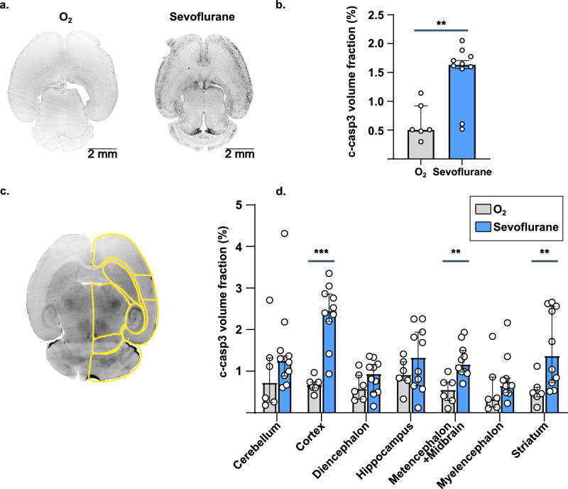
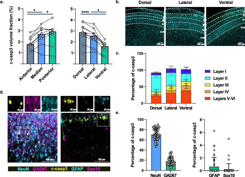
In the last two decades, safety concerns about general anesthesia (GA) arose from studies documenting brain cell death in various pharmacological conditions and animal models. Nowadays, a thorough characterization of sevoflurane-induced apoptosis in the entire neonatal mouse brain would help identify and further focus on underlying mechanisms. We performed whole-brain mapping of sevoflurane-induced apoptosis in post-natal day (P) 7 mice using tissue clearing and immunohistochemistry. We found an anatomically heterogenous increase in cleaved-caspase-3 staining. The use of a novel P7 brain atlas showed that the neocortex was the most affected area, followed by the striatum and the metencephalon. Histological characterization in cortical slices determined that post-mitotic neurons were the most affected cell type and followed inter- and intracortical gradients with maximal apoptosis in the superficial layers of the posterodorsal cortex. The unbiased anatomical mapping used here allowed us to confirm sevoflurane-induced apoptosis in the perinatal period, neocortical involvement, and indicated striatal and metencephalic damage while suggesting moderate hippocampal one. The identification of neocortical gradients is consistent with a maturity-dependent mechanism. Further research could then focus on the interference of sevoflurane with neuronal migration and survival during development.
在过去的二十年中,由于研究记录了各种药理学条件和动物模型中的脑细胞死亡,人们对全身麻醉(GA)的安全性产生了担忧。如今,对新生小鼠大脑中七氟醚诱导的细胞凋亡进行全面描述将有助于确定和进一步关注潜在的机制。我们使用组织透明化和免疫组织化学技术对出生后第 7 天(P)的小鼠进行了全脑映射,以检测七氟醚诱导的凋亡。我们发现,在解剖学上存在异质性的 caspase-3 裂解染色增加。使用新的 P7 大脑图谱显示,新皮质是受影响最严重的区域,其次是纹状体和后脑。皮质切片的组织学特征确定,有丝分裂后神经元是最易受影响的细胞类型,随后是皮质内和皮质间梯度,在后背侧皮质的浅层凋亡达到最大值。这里使用的无偏的解剖学映射方法使我们能够确认围产期七氟醚诱导的细胞凋亡、新皮质的参与,并表明纹状体和后脑的损伤,同时提示海马体的损伤程度适中。新皮质梯度的确定与成熟依赖性机制一致。进一步的研究可以集中在七氟醚对发育过程中神经元迁移和存活的干扰上。