Schmidt Gilda, Findeklee Sebastian, Del Sol Martinez Gerda, Georgescu Mihai-Teodor, Gerlinger Christoph, Nemat Sogand, Klamminger Gilbert Georg, Nigdelis Meletios P, Solomayer Erich-Franz, Hamoud Bashar Haj
Department for Gynecology, Obstetrics and Reproductive Medicine, Saarland University Hospital, 66421 Homburg, Germany.
"Prof. Dr. Al. Trestioreanu" Oncology Discipline, "Carol Davila" University of Medicine and Pharmacy, 020021 Bucharest, Romania.
Diagnostics (Basel). 2023 Aug 30;13(17):2811. doi: 10.3390/diagnostics13172811.
Nowadays chemotherapy in breast cancer patients is optionally applied neoadjuvant, which allows for testing of tumor response to the chemotherapeutical treatment in vivo, as well as allowing a greater number of patients to benefit from a subsequent breast-conserving surgery.
We compared breast ultrasonography, mammography, and clinical examination (palpation) results with postoperative histopathological findings after neoadjuvant chemotherapy, aiming to determine the most accurate prediction of complete remission and tumor-free resection margins. To this end, clinical and imaging data of 184 patients (193 tumors) with confirmed diagnosis of breast cancer and neoadjuvant therapy were analyzed.
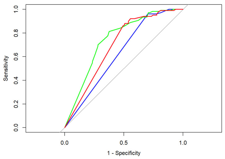
After chemotherapy, tumors could be assessed by palpation in 91.7%, by sonography in 99.5%, and by mammography in 84.5% (chi-square < 0.0001) of cases. Although mammography proved more accurate in estimating the exact neoadjuvant tumor size than breast sonography in total numbers (136/163 (83.44%) vs. 142/192 (73.96%), n.s.), 29 tumors could be assessed solely by means of breast sonography. A sonographic measurement was feasible in 192 cases (99.48%) post-chemotherapy and in all cases prior to chemotherapy.
We determined a superiority of mammography and breast sonography over clinical palpation in predicting neoadjuvant tumor size. However, neither examination method can predict either pCR or tumor margins with high confidence.
如今,乳腺癌患者的化疗可选择新辅助化疗,这使得能够在体内测试肿瘤对化疗治疗的反应,同时也让更多患者能够受益于后续的保乳手术。
我们将新辅助化疗后乳房超声检查、乳腺X线摄影和临床检查(触诊)结果与术后组织病理学结果进行比较,旨在确定对完全缓解和无瘤切除边缘的最准确预测。为此,分析了184例(193个肿瘤)确诊为乳腺癌并接受新辅助治疗患者的临床和影像学数据。
化疗后,91.7%的病例可通过触诊评估肿瘤,99.5%可通过超声检查评估,84.5%可通过乳腺X线摄影评估(卡方检验<0.0001)。尽管总体上乳腺X线摄影在估计新辅助化疗后肿瘤的确切大小方面比乳房超声检查更准确(136/163(83.44%)对142/192(73.96%),无统计学差异),但有29个肿瘤只能通过乳房超声检查评估。化疗后192例(99.48%)可行超声测量,化疗前所有病例均可行。
我们确定在预测新辅助化疗后肿瘤大小方面,乳腺X线摄影和乳房超声检查优于临床触诊。然而,两种检查方法都不能高度准确地预测病理完全缓解或肿瘤边缘。