Chiu James, Bova Davide, Spear Georgia, Ecanow Jacob, Choate Alyssa, Besson Pierre, Caluser Calin
Department of Radiology, Endeavor Health, 2650 Ridge Ave, Evanston, IL 60201, USA.
Dacia Medical Clinic, 917 S Oak Park Ave, Suite B, Oak Park, IL 60304, USA.
Diagnostics (Basel). 2024 Jul 25;14(15):1602. doi: 10.3390/diagnostics14151602.
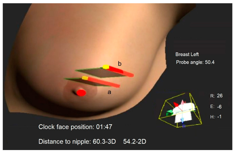
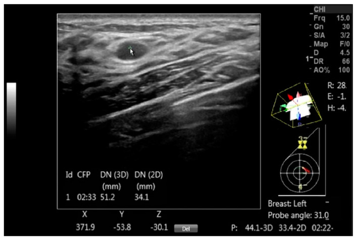
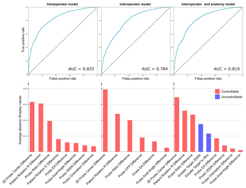
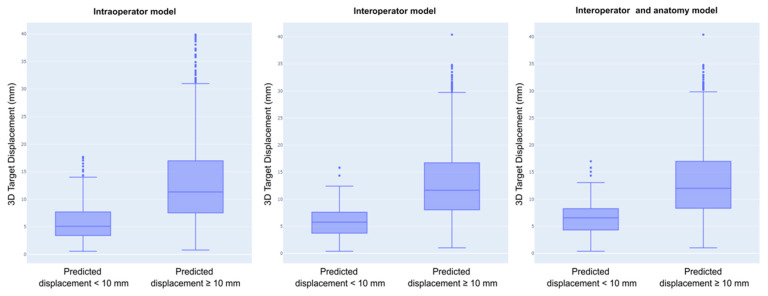
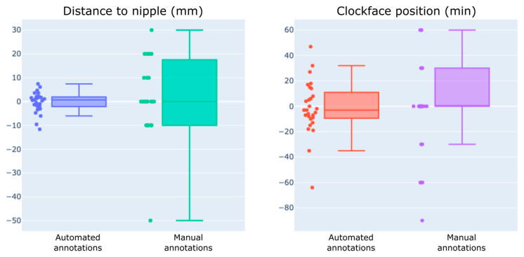
Interoperator variability in the reproducibility of breast lesions found by handheld ultrasound (HHUS) can significantly interfere with clinical care. This study analyzed the features associated with breast mass position differences during HHUS. The ability of operators to reproduce the position of small masses and the time required to generate annotations with and without a computer-assisted scanning device (DEVICE) were also evaluated. This prospective study included 28 patients with 34 benign or probably benign small breast masses. Two operators generated manual and automated position annotations for each mass. The probe and body positions were systematically varied during scanning with the DEVICE, and the features describing mass movement were used in three logistic regression models trained to discriminate small from large breast mass displacements (cutoff: 10 mm). All models successfully discriminated small from large breast mass displacements (areas under the curve: 0.78 to 0.82). The interoperator localization precision was 6.6 ± 2.8 mm with DEVICE guidance and 19.9 ± 16.1 mm with manual annotations. Computer-assisted scanning reduced the time to annotate and reidentify a mass by 33 and 46 s on average, respectively. The results demonstrated that breast mass location reproducibility and exam efficiency improved by controlling operator actionable features with computer-assisted HHUS.
手持式超声(HHUS)检查发现的乳腺病变再现性方面的操作者间差异会显著干扰临床护理。本研究分析了HHUS检查期间与乳腺肿块位置差异相关的特征。还评估了操作者再现小肿块位置的能力以及使用和不使用计算机辅助扫描设备(DEVICE)生成标注所需的时间。这项前瞻性研究纳入了28例患者,其乳房有34个良性或可能为良性的小肿块。两名操作者为每个肿块生成手动和自动位置标注。在使用DEVICE扫描期间,探头和身体位置系统地变化,描述肿块移动的特征被用于三个逻辑回归模型,这些模型经过训练以区分小乳腺肿块和大乳腺肿块的位移(临界值:10毫米)。所有模型均成功区分了小乳腺肿块和大乳腺肿块的位移(曲线下面积:0.78至0.82)。在DEVICE引导下,操作者间的定位精度为6.6±2.8毫米,手动标注时为19.9±16.1毫米。计算机辅助扫描平均分别将标注和重新识别肿块的时间减少了33秒和46秒。结果表明,通过计算机辅助HHUS控制操作者可操作的特征,可提高乳腺肿块位置的再现性和检查效率。