• 文献检索
  • 文档翻译
  • 深度研究
  • 学术资讯
  • Suppr Zotero 插件Zotero 插件
  • 邀请有礼
  • 套餐&价格
  • 历史记录
应用&插件
Suppr Zotero 插件Zotero 插件浏览器插件Mac 客户端Windows 客户端微信小程序
定价
高级版会员购买积分包购买API积分包
服务
文献检索文档翻译深度研究API 文档MCP 服务
关于我们
关于 Suppr公司介绍联系我们用户协议隐私条款
关注我们

Suppr 超能文献

核心技术专利:CN118964589B侵权必究
粤ICP备2023148730 号-1Suppr @ 2026

文献检索

告别复杂PubMed语法,用中文像聊天一样搜索,搜遍4000万医学文献。AI智能推荐,让科研检索更轻松。

立即免费搜索

文件翻译

保留排版,准确专业,支持PDF/Word/PPT等文件格式,支持 12+语言互译。

免费翻译文档

深度研究

AI帮你快速写综述,25分钟生成高质量综述,智能提取关键信息,辅助科研写作。

立即免费体验

突变体中突触内质网腔蛋白容纳的破坏

Disruption of Synaptic Endoplasmic Reticulum Luminal Protein Containment in Mutants.

作者信息

Quiñones-Frías Mónica C, Ocken Dina M, Rodal Avital

机构信息

Department of Biology, Brandeis University, Waltham, MA.

出版信息

bioRxiv. 2025 Feb 15:2023.09.01.555994. doi: 10.1101/2023.09.01.555994.

DOI:10.1101/2023.09.01.555994
PMID:37693578
原文链接:https://pmc.ncbi.nlm.nih.gov/articles/PMC10491308/
Abstract

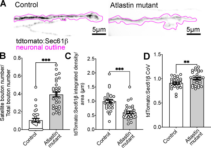
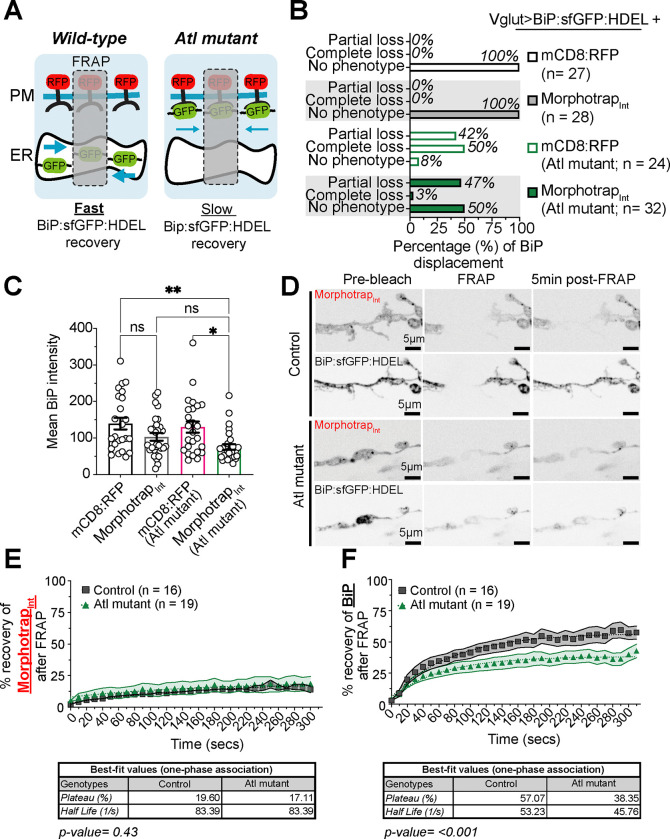
The endoplasmic reticulum (ER) extends throughout neurons and regulates many neuronal functions, including neurite outgrowth, neurotransmission, and synaptic plasticity. Mutations in proteins that control ER shape are linked to the neurodegenerative disorder Hereditary Spastic Paraplegia (HSP), yet the ultrastructure and dynamics of neuronal ER remain largely unexplored, especially at presynaptic terminals. Using super-resolution and live imaging in larval motor neurons, we investigated ER structure at presynaptic terminals of wild-type animals and null mutants of the ER shaping protein and HSP-linked gene, Atlastin. Previous studies using an ER luminal marker reported diffuse localization at mutant presynaptic terminals, which was attributed to ER fragmentation. However, using an ER membrane marker, we discovered that mutant ER forms robust networks with only mild defects in structural dynamics, indicating the primary defect is functional rather than architectural. We demonstrate that mutants progressively displace overexpressed luminal ER proteins to the cytosol during larval development, specifically at synapses, while these proteins remain correctly localized in cell bodies, axons, and muscles. This synaptic-specific displacement phenotype, previously unreported in non-neuronal cells, emphasizes the importance of studying neurons to understand HSP pathogenesis.

摘要

内质网(ER)贯穿神经元并调节许多神经元功能,包括神经突生长、神经传递和突触可塑性。控制内质网形态的蛋白质突变与神经退行性疾病遗传性痉挛性截瘫(HSP)有关,但神经元内质网的超微结构和动力学在很大程度上仍未得到探索,尤其是在突触前终末。利用幼虫运动神经元的超分辨率和实时成像技术,我们研究了野生型动物以及内质网塑形蛋白和与HSP相关基因Atlastin的无效突变体的突触前终末的内质网结构。先前使用内质网腔标记物的研究报道,在突变体突触前终末内质网呈弥漫性定位,这被归因于内质网碎片化。然而,使用内质网膜标记物,我们发现突变体的内质网形成了强大的网络,结构动力学仅存在轻微缺陷,这表明主要缺陷是功能性的而非结构性的。我们证明,在幼虫发育过程中,特别是在突触处,突变体逐渐将过表达的内质网腔蛋白转移到细胞质中,而这些蛋白在细胞体、轴突和肌肉中仍正确定位。这种突触特异性转移表型,此前在非神经元细胞中未被报道,强调了研究神经元对于理解HSP发病机制的重要性。

https://cdn.ncbi.nlm.nih.gov/pmc/blobs/a6e1/11867439/8f549255b7cf/nihpp-2023.09.01.555994v2-f0014.jpg
https://cdn.ncbi.nlm.nih.gov/pmc/blobs/a6e1/11867439/84304775f987/nihpp-2023.09.01.555994v2-f0001.jpg
https://cdn.ncbi.nlm.nih.gov/pmc/blobs/a6e1/11867439/77f63095e8f8/nihpp-2023.09.01.555994v2-f0002.jpg
https://cdn.ncbi.nlm.nih.gov/pmc/blobs/a6e1/11867439/57a8b0717e61/nihpp-2023.09.01.555994v2-f0003.jpg
https://cdn.ncbi.nlm.nih.gov/pmc/blobs/a6e1/11867439/f3d41e6ff963/nihpp-2023.09.01.555994v2-f0004.jpg
https://cdn.ncbi.nlm.nih.gov/pmc/blobs/a6e1/11867439/a35cb4c1846c/nihpp-2023.09.01.555994v2-f0005.jpg
https://cdn.ncbi.nlm.nih.gov/pmc/blobs/a6e1/11867439/e81a811212b2/nihpp-2023.09.01.555994v2-f0006.jpg
https://cdn.ncbi.nlm.nih.gov/pmc/blobs/a6e1/11867439/c123d454a649/nihpp-2023.09.01.555994v2-f0007.jpg
https://cdn.ncbi.nlm.nih.gov/pmc/blobs/a6e1/11867439/cf471e68fbf2/nihpp-2023.09.01.555994v2-f0008.jpg
https://cdn.ncbi.nlm.nih.gov/pmc/blobs/a6e1/11867439/766b37946729/nihpp-2023.09.01.555994v2-f0009.jpg
https://cdn.ncbi.nlm.nih.gov/pmc/blobs/a6e1/11867439/60d27ca54ac1/nihpp-2023.09.01.555994v2-f0010.jpg
https://cdn.ncbi.nlm.nih.gov/pmc/blobs/a6e1/11867439/58e7d659f2b8/nihpp-2023.09.01.555994v2-f0011.jpg
https://cdn.ncbi.nlm.nih.gov/pmc/blobs/a6e1/11867439/f6165cae6408/nihpp-2023.09.01.555994v2-f0012.jpg
https://cdn.ncbi.nlm.nih.gov/pmc/blobs/a6e1/11867439/82b29424c2c6/nihpp-2023.09.01.555994v2-f0013.jpg
https://cdn.ncbi.nlm.nih.gov/pmc/blobs/a6e1/11867439/8f549255b7cf/nihpp-2023.09.01.555994v2-f0014.jpg

相似文献

1
Disruption of Synaptic Endoplasmic Reticulum Luminal Protein Containment in Mutants.突变体中突触内质网腔蛋白容纳的破坏
bioRxiv. 2025 Feb 15:2023.09.01.555994. doi: 10.1101/2023.09.01.555994.
2
Atlastin in motor neurons is required for locomotion and presynaptic function.在运动神经元中,Atlastin 的缺失会导致运动和突触前功能异常。
J Cell Sci. 2017 Oct 15;130(20):3507-3516. doi: 10.1242/jcs.201657. Epub 2017 Aug 31.
3
Endoplasmic Reticulum Lumenal Indicators in Reveal Effects of HSP-Related Mutations on Endoplasmic Reticulum Calcium Dynamics.内质网腔标志物揭示热休克蛋白相关突变对内质网钙动力学的影响。
Front Neurosci. 2020 Aug 10;14:816. doi: 10.3389/fnins.2020.00816. eCollection 2020.
4
The effects of ER morphology on synaptic structure and function in Drosophila melanogaster.内质网形态对黑腹果蝇突触结构和功能的影响。
J Cell Sci. 2016 Apr 15;129(8):1635-48. doi: 10.1242/jcs.184929. Epub 2016 Feb 23.
5
Drosophila SPG12 ortholog, reticulon-like 1, governs presynaptic ER organization and Ca2+ dynamics.果蝇 SPG12 同源物,网质蛋白样 1,调节突触前内质网的组织和 Ca2+动力学。
J Cell Biol. 2023 Jun 5;222(6). doi: 10.1083/jcb.202112101. Epub 2023 Mar 23.
6
Hereditary spastic paraplegia proteins REEP1, spastin, and atlastin-1 coordinate microtubule interactions with the tubular ER network.遗传性痉挛性截瘫蛋白 REEP1、spastin 和 atlastin-1 协调微管与管状内质网网络的相互作用。
J Clin Invest. 2010 Apr;120(4):1097-110. doi: 10.1172/JCI40979.
7
Atlastin-1 regulates endosomal tubulation and lysosomal proteolysis in human cortical neurons.Atlastin-1 调节人皮质神经元内体小管化和溶酶体蛋白水解。
Neurobiol Dis. 2024 Sep;199:106556. doi: 10.1016/j.nbd.2024.106556. Epub 2024 Jun 6.
8
Atlastin regulates store-operated calcium entry for nerve growth factor-induced neurite outgrowth.Atlastin 调节钙库操纵型钙内流,促进神经营养因子诱导的轴突生长。
Sci Rep. 2017 Feb 27;7:43490. doi: 10.1038/srep43490.
9
Atlastin-1 regulates morphology and function of endoplasmic reticulum in dendrites.Atlastin-1 调节树突内质网的形态和功能。
Nat Commun. 2019 Feb 4;10(1):568. doi: 10.1038/s41467-019-08478-6.
10
Fusing a lasting relationship between ER tubules.将内质网小管融合在一起。
Trends Cell Biol. 2011 Jul;21(7):416-23. doi: 10.1016/j.tcb.2011.03.009. Epub 2011 May 6.

本文引用的文献

1
Periodic ER-plasma membrane junctions support long-range Ca signal integration in dendrites.周期性内质网-质膜连接支持树突中的长距离钙信号整合。
Cell. 2025 Jan 23;188(2):484-500.e22. doi: 10.1016/j.cell.2024.11.029. Epub 2024 Dec 20.
2
The membrane curvature-inducing REEP1-4 proteins generate an ER-derived vesicular compartment.REEP1-4 蛋白诱导的膜弯曲生成了一个源自内质网的囊泡隔室。
Nat Commun. 2024 Oct 5;15(1):8655. doi: 10.1038/s41467-024-52901-6.
3
Luminal transport through intact endoplasmic reticulum limits the magnitude of localized Ca signals.
内质网腔运输通过完整的内质网限制局部 Ca 信号的幅度。
Proc Natl Acad Sci U S A. 2024 Mar 26;121(13):e2312172121. doi: 10.1073/pnas.2312172121. Epub 2024 Mar 19.
4
Motion of VAPB molecules reveals ER-mitochondria contact site subdomains.VAPB 分子的运动揭示了内质网-线粒体接触位点亚区。
Nature. 2024 Feb;626(7997):169-176. doi: 10.1038/s41586-023-06956-y. Epub 2024 Jan 24.
5
Mechanisms of substrate processing during ER-associated protein degradation.内质网相关蛋白降解过程中底物加工的机制。
Nat Rev Mol Cell Biol. 2023 Nov;24(11):777-796. doi: 10.1038/s41580-023-00633-8. Epub 2023 Aug 1.
6
Drosophila motor neuron boutons remodel through membrane blebbing coupled with muscle contraction.果蝇运动神经元末梢通过与肌肉收缩偶联的细胞膜起泡进行重塑。
Nat Commun. 2023 Jun 8;14(1):3352. doi: 10.1038/s41467-023-38421-9.
7
Drosophila SPG12 ortholog, reticulon-like 1, governs presynaptic ER organization and Ca2+ dynamics.果蝇 SPG12 同源物,网质蛋白样 1,调节突触前内质网的组织和 Ca2+动力学。
J Cell Biol. 2023 Jun 5;222(6). doi: 10.1083/jcb.202112101. Epub 2023 Mar 23.
8
An approach for quantitative mapping of synaptic periactive zone architecture and organization.一种定量映射突触活性区结构和组织的方法。
Mol Biol Cell. 2023 May 15;34(6):ar51. doi: 10.1091/mbc.E22-08-0372. Epub 2022 Dec 21.
9
Endoplasmic Reticulum Stress Signaling and Neuronal Cell Death.内质网应激信号与神经元细胞死亡。
Int J Mol Sci. 2022 Dec 2;23(23):15186. doi: 10.3390/ijms232315186.
10
The ER ladder is a unique morphological feature of developing mammalian axons.ER 梯是发育中的哺乳动物轴突的独特形态特征。
Dev Cell. 2022 Jun 6;57(11):1369-1382.e6. doi: 10.1016/j.devcel.2022.05.002. Epub 2022 May 23.