• 文献检索
  • 文档翻译
  • 深度研究
  • 学术资讯
  • Suppr Zotero 插件Zotero 插件
  • 邀请有礼
  • 套餐&价格
  • 历史记录
应用&插件
Suppr Zotero 插件Zotero 插件浏览器插件Mac 客户端Windows 客户端微信小程序
定价
高级版会员购买积分包购买API积分包
服务
文献检索文档翻译深度研究API 文档MCP 服务
关于我们
关于 Suppr公司介绍联系我们用户协议隐私条款
关注我们

Suppr 超能文献

核心技术专利:CN118964589B侵权必究
粤ICP备2023148730 号-1Suppr @ 2026

文献检索

告别复杂PubMed语法,用中文像聊天一样搜索,搜遍4000万医学文献。AI智能推荐,让科研检索更轻松。

立即免费搜索

文件翻译

保留排版,准确专业,支持PDF/Word/PPT等文件格式,支持 12+语言互译。

免费翻译文档

深度研究

AI帮你快速写综述,25分钟生成高质量综述,智能提取关键信息,辅助科研写作。

立即免费体验

无症状的杜氏肌营养不良基因突变女性携带者的心肌固有 T 映射和细胞外容积定量。

Myocardial native T mapping and extracellular volume quantification in asymptomatic female carriers of Duchenne muscular dystrophy gene mutations.

机构信息

International Clinical Research Centre, St. Anne's University Hospital, Brno, Czech Republic.

1st Department of Internal Medicine-Cardioangiology, St. Anne's University Hospital, Faculty of Medicine, Masaryk University, Brno, Czech Republic.

出版信息

Orphanet J Rare Dis. 2023 Sep 11;18(1):283. doi: 10.1186/s13023-023-02899-9.

DOI:10.1186/s13023-023-02899-9
PMID:37697356
原文链接:https://pmc.ncbi.nlm.nih.gov/articles/PMC10496385/
Abstract

BACKGROUND

Female carriers of dystrophin gene mutations (DMD-FC) were previously considered non-manifesting, but in recent decades, cardiomyopathy associated with muscular dystrophy and myocardial fibrosis has been described. Our study aimed to assess prospectively myocardial fibrosis in asymptomatic DMD-FC compared to a sex-matched control group (CG) with similar age distribution using native T mapping and extracellular volume (ECV) quantification by cardiovascular magnetic resonance (CMR) imaging.

MATERIALS AND METHODS

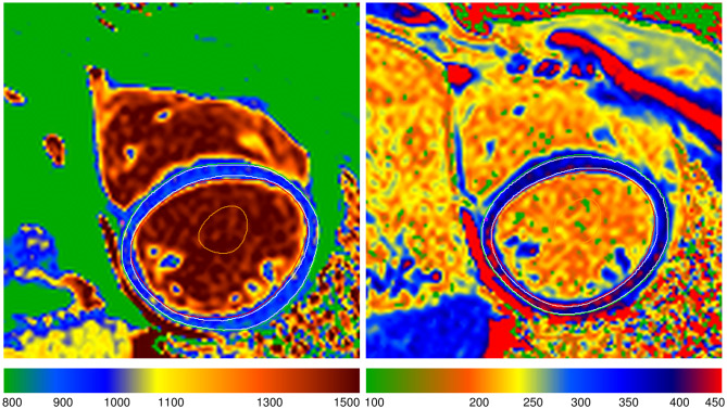
38 DMD-FC with verified genetic mutation and 22 healthy volunteers were included. Using CMR, native T relaxation time and ECV quantification were determined in each group. Late gadolinium enhancement (LGE) was assessed in all cases.

RESULTS

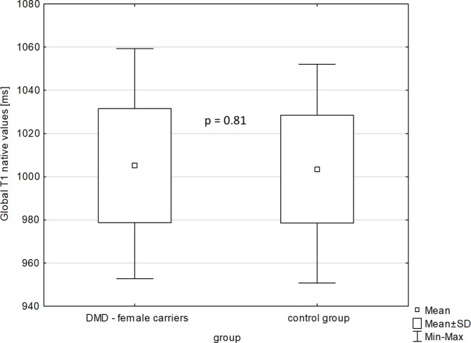
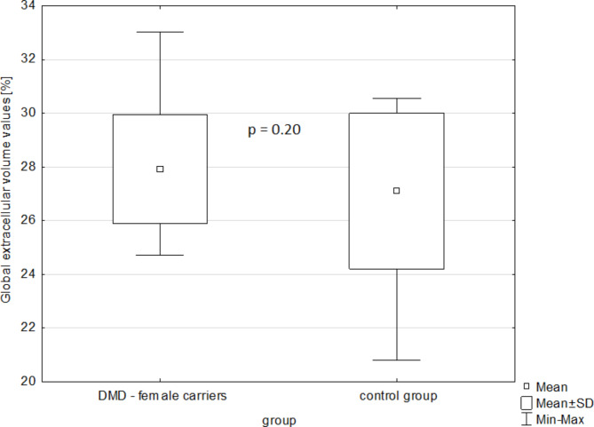
There were 38 DMD-FC (mean age 39.1 ± 8.8 years) and 22 healthy volunteers (mean age 39.9 ± 12.6 years) imagined by CMR. The mean global native T relaxation time was similar for DMD-FC and CG (1005.1 ± 26.3 ms vs. 1003.5 ± 25.0 ms; p-value = 0.81). Likewise, the mean global ECV value was also similar between the groups (27.92 ± 2.02% vs. 27.10 ± 2.89%; p-value = 0.20). The segmental analysis of mean ECV values according to the American Heart Association classification did not show any differences between DMD-FC and CG. There was a non-significant trend towards higher mean ECV values of DMD-FC in the inferior and inferolateral segments of the myocardium (p-value = 0.075 and 0.070 respectively).

CONCLUSION

There were no statistically significant differences in the mean global and segmental native T relaxation times and the mean global or segmental ECV values. There was a trend towards higher segmental mean ECV values of DMD-FC in the inferior and inferolateral walls of the myocardium.

摘要

背景

肌营养不良蛋白基因突变的女性携带者(DMD-FC)以前被认为是非表现型的,但在最近几十年,与肌肉营养不良相关的心肌病和心肌纤维化已被描述。我们的研究旨在使用心血管磁共振(CMR)成像的原生 T 映射和细胞外体积(ECV)定量,前瞻性地评估无症状 DMD-FC 与具有相似年龄分布的性别匹配对照组(CG)的心肌纤维化。

材料和方法

38 名经基因验证突变的 DMD-FC 和 22 名健康志愿者被纳入研究。在每组中,使用 CMR 确定原生 T 弛豫时间和 ECV 定量。在所有病例中评估晚期钆增强(LGE)。

结果

38 名 DMD-FC(平均年龄 39.1±8.8 岁)和 22 名健康志愿者(平均年龄 39.9±12.6 岁)接受了 CMR 成像。DMD-FC 和 CG 的平均整体原生 T 弛豫时间相似(1005.1±26.3 ms 与 1003.5±25.0 ms;p 值=0.81)。同样,两组的平均整体 ECV 值也相似(27.92±2.02%与 27.10±2.89%;p 值=0.20)。根据美国心脏协会分类的平均 ECV 值的节段分析显示,DMD-FC 和 CG 之间没有差异。DMD-FC 的下壁和下侧壁心肌的平均 ECV 值有升高的非显著趋势(p 值分别为 0.075 和 0.070)。

结论

平均整体和节段性原生 T 弛豫时间以及平均整体或节段性 ECV 值没有统计学差异。DMD-FC 的下壁和下侧壁心肌节段性平均 ECV 值有升高的趋势。

https://cdn.ncbi.nlm.nih.gov/pmc/blobs/f275/10496385/988be6ff36f2/13023_2023_2899_Fig4_HTML.jpg
https://cdn.ncbi.nlm.nih.gov/pmc/blobs/f275/10496385/28d56018bf4f/13023_2023_2899_Fig1_HTML.jpg
https://cdn.ncbi.nlm.nih.gov/pmc/blobs/f275/10496385/2b489f7bad84/13023_2023_2899_Fig2_HTML.jpg
https://cdn.ncbi.nlm.nih.gov/pmc/blobs/f275/10496385/732011bf9c48/13023_2023_2899_Fig3_HTML.jpg
https://cdn.ncbi.nlm.nih.gov/pmc/blobs/f275/10496385/988be6ff36f2/13023_2023_2899_Fig4_HTML.jpg

相似文献

1
Myocardial native T mapping and extracellular volume quantification in asymptomatic female carriers of Duchenne muscular dystrophy gene mutations.无症状的杜氏肌营养不良基因突变女性携带者的心肌固有 T 映射和细胞外容积定量。
Orphanet J Rare Dis. 2023 Sep 11;18(1):283. doi: 10.1186/s13023-023-02899-9.
2
T-Mapping and extracellular volume estimates in pediatric subjects with Duchenne muscular dystrophy and healthy controls at 3T.3T 下杜氏肌营养不良症患儿与健康对照者的 T 映射和细胞外容积评估。
J Cardiovasc Magn Reson. 2020 Dec 10;22(1):85. doi: 10.1186/s12968-020-00687-z.
3
Increased myocardial native T1 and extracellular volume in patients with Duchenne muscular dystrophy.杜兴氏肌营养不良症患者心肌固有T1和细胞外容积增加。
J Cardiovasc Magn Reson. 2016 Jan 21;18:5. doi: 10.1186/s12968-016-0224-7.
4
Native T1 values identify myocardial changes and stratify disease severity in patients with Duchenne muscular dystrophy.固有T1值可识别杜氏肌营养不良症患者的心肌变化并对疾病严重程度进行分层。
J Cardiovasc Magn Reson. 2016 Oct 28;18(1):72. doi: 10.1186/s12968-016-0292-8.
5
Elevated Myocardial Extracellular Volume Fraction in Duchenne Muscular Dystrophy.杜氏肌营养不良症中心肌细胞外容积分数升高
Pediatr Cardiol. 2017 Oct;38(7):1485-1492. doi: 10.1007/s00246-017-1690-x. Epub 2017 Jul 31.
6
Global, segmental and layer specific analysis of myocardial involvement in Duchenne muscular dystrophy by cardiovascular magnetic resonance native T1 mapping.应用心血管磁共振 native T1 mapping 对杜氏肌营养不良症心肌受累的全局、节段和分层分析。
J Cardiovasc Magn Reson. 2021 Oct 14;23(1):110. doi: 10.1186/s12968-021-00802-8.
7
Cardiac profile of the Czech population of Duchenne muscular dystrophy patients: a cardiovascular magnetic resonance study with T1 mapping.捷克杜氏肌营养不良症患者的心脏特征:T1 映射的心血管磁共振研究。
Orphanet J Rare Dis. 2019 Jan 9;14(1):10. doi: 10.1186/s13023-018-0986-0.
8
Myocardial fibrosis imaging based on T1-mapping and extracellular volume fraction (ECV) measurement in muscular dystrophy patients: diagnostic value compared with conventional late gadolinium enhancement (LGE) imaging.基于T1映射和细胞外容积分数(ECV)测量的肌营养不良患者心肌纤维化成像:与传统钆延迟增强(LGE)成像相比的诊断价值
Eur Heart J Cardiovasc Imaging. 2014 Sep;15(9):1004-12. doi: 10.1093/ehjci/jeu050. Epub 2014 Mar 30.
9
Comprehensive cardiac magnetic resonance T1, T2, and extracellular volume mapping to define Duchenne cardiomyopathy.综合心脏磁共振 T1、T2 和细胞外容积成像用于定义杜氏肌营养不良症的心肌病变。
J Cardiovasc Magn Reson. 2023 Jul 31;25(1):44. doi: 10.1186/s12968-023-00951-y.
10
Non-contrast cardiovascular magnetic resonance detection of myocardial fibrosis in Duchenne muscular dystrophy.磁共振非增强扫描检测杜氏肌营养不良症心肌纤维化。
J Cardiovasc Magn Reson. 2021 Apr 29;23(1):48. doi: 10.1186/s12968-021-00736-1.

引用本文的文献

1
Heart Disease in Mothers of Children with Duchenne Muscular Dystrophy.患有杜氏肌营养不良症患儿的母亲的心脏病。
Curr Cardiol Rev. 2024;20(6):e230724232195. doi: 10.2174/011573403X292850240719074112.
2
Contrast-Enhanced Magnetic Resonance Imaging Based T1 Mapping and Extracellular Volume Fractions Are Associated with Peripheral Artery Disease.基于对比增强磁共振成像的T1映射和细胞外体积分数与外周动脉疾病相关。
J Cardiovasc Dev Dis. 2024 Jun 14;11(6):181. doi: 10.3390/jcdd11060181.
3
Asymmetric Myocardial Involvement as an Early Indicator of Cardiac Dysfunction in Pediatric Dystrophinopathies: A Study on Cardiac Magnetic Resonance (CMR) Parametric Mappings.

本文引用的文献

1
The clinical and prognostic value of late Gadolinium enhancement imaging in heart failure with mid-range and preserved ejection fraction.心力衰竭伴中间范围和射血分数保留患者中晚期钆增强成像的临床和预后价值。
Heart Vessels. 2022 Feb;37(2):273-281. doi: 10.1007/s00380-021-01910-2. Epub 2021 Jul 22.
2
Quantitative assessment of left ventricular longitudinal function and myocardial deformation in Duchenne muscular dystrophy patients.定量评估杜氏肌营养不良症患者左心室纵向功能和心肌变形。
Orphanet J Rare Dis. 2021 Jan 30;16(1):57. doi: 10.1186/s13023-021-01704-9.
3
Echocardiographic signs of subclinical cardiac function impairment in Duchenne dystrophy gene carriers.
不对称性心肌受累作为小儿肌营养不良症心脏功能障碍的早期指标:心脏磁共振(CMR)参数映射研究
Pediatr Cardiol. 2025 Mar;46(3):685-693. doi: 10.1007/s00246-024-03488-8. Epub 2024 Apr 30.
4
Correction: Myocardial native T mapping and extracellular volume quantification in asymptomatic female carriers of Duchenne muscular dystrophy gene mutations.更正:杜兴氏肌营养不良基因突变无症状女性携带者的心肌天然T映射和细胞外容积定量分析
Orphanet J Rare Dis. 2023 Oct 19;18(1):331. doi: 10.1186/s13023-023-02922-z.
超声心动图在杜氏肌营养不良症基因突变携带者亚临床心功能障碍中的表现
Sci Rep. 2020 Nov 27;10(1):20794. doi: 10.1038/s41598-020-77882-6.
4
Decreased Global Strains of LV in Asymptomatic Female Duchenne Muscular Dystrophy Gene Carriers Using CMR-FT.使用心脏磁共振标记成像(CMR-FT)检测无症状女性杜氏肌营养不良基因携带者左心室整体应变降低
JACC Cardiovasc Imaging. 2021 May;14(5):1070-1072. doi: 10.1016/j.jcmg.2020.09.016. Epub 2020 Nov 18.
5
Duchenne and Becker muscular dystrophy carriers: Evidence of cardiomyopathy by exercise and cardiac MRI testing.杜兴氏和贝克氏肌营养不良症携带者:通过运动和心脏磁共振成像测试发现心肌病的证据。
Int J Cardiol. 2020 Oct 1;316:257-265. doi: 10.1016/j.ijcard.2020.05.052. Epub 2020 May 27.
6
Progressive left ventricular dysfunction and myocardial fibrosis in Duchenne and Becker muscular dystrophy: a longitudinal cardiovascular magnetic resonance study.杜兴氏和贝克氏肌营养不良症患者的进行性左心室功能障碍和心肌纤维化:一项纵向心血管磁共振研究。
Pediatr Cardiol. 2019 Feb;40(2):384-392. doi: 10.1007/s00246-018-2046-x. Epub 2018 Dec 18.
7
Management of Cardiac Involvement Associated With Neuromuscular Diseases: A Scientific Statement From the American Heart Association.与神经肌肉疾病相关的心脏管理:美国心脏协会的科学声明。
Circulation. 2017 Sep 26;136(13):e200-e231. doi: 10.1161/CIR.0000000000000526. Epub 2017 Aug 24.
8
Elevated Myocardial Extracellular Volume Fraction in Duchenne Muscular Dystrophy.杜氏肌营养不良症中心肌细胞外容积分数升高
Pediatr Cardiol. 2017 Oct;38(7):1485-1492. doi: 10.1007/s00246-017-1690-x. Epub 2017 Jul 31.
9
Native T1 values identify myocardial changes and stratify disease severity in patients with Duchenne muscular dystrophy.固有T1值可识别杜氏肌营养不良症患者的心肌变化并对疾病严重程度进行分层。
J Cardiovasc Magn Reson. 2016 Oct 28;18(1):72. doi: 10.1186/s12968-016-0292-8.
10
Cardiac involvement in female carriers of duchenne or becker muscular dystrophy.杜兴氏或贝克氏肌营养不良女性携带者的心脏受累情况。
Muscle Nerve. 2017 Jun;55(6):810-818. doi: 10.1002/mus.25445. Epub 2017 Feb 15.