Department of Neuroscience (DNS), University of Padova, Via Giustiniani 5, 35128, Padua, Italy.
Padova Neuroscience Center, University of Padova, Padua, Italy.
J Neurol. 2024 Jan;271(1):300-309. doi: 10.1007/s00415-023-11962-1. Epub 2023 Sep 12.
To investigate brain MRI abnormalities in a cohort of patients with rapidly progressive dementia (RPD) with and without a diagnosis of Creutzfeldt-Jakob disease (CJD).
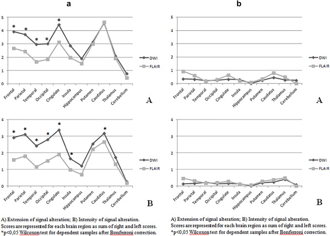
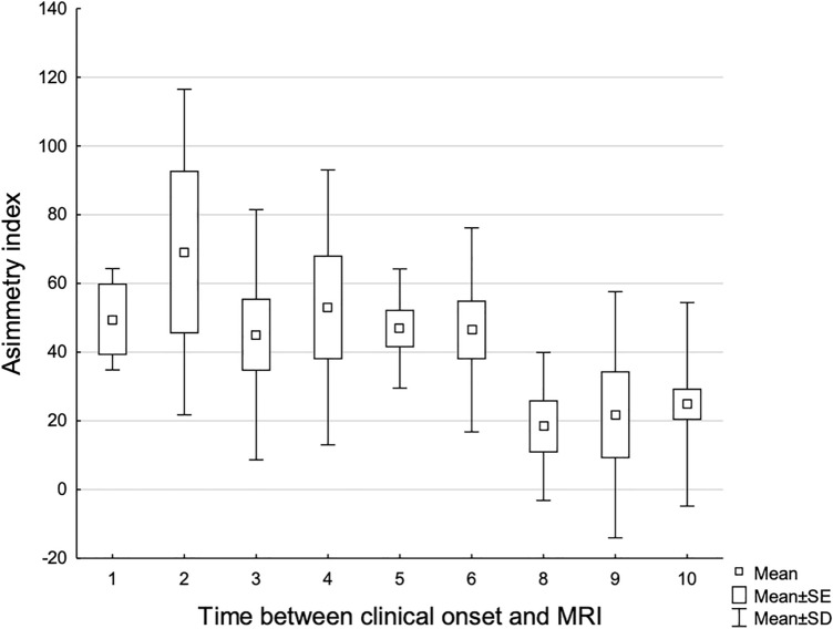
One hundred and seven patients with diagnosis of prion disease (60 with definite sCJD, 33 with probable sCJD and 14 with genetic prion disease) and 40 non-prion related RPD patients (npRPD) underwent brain MRI including DWI and FLAIR. MRIs were evaluated with a semiquantitative rating score, which separately considered abnormal signal extent and intensity in 22 brain regions. Clinical findings at onset, disease duration, cerebrospinal-fluid 14-3-3 and t-tau protein levels, and EEG data were recorded.
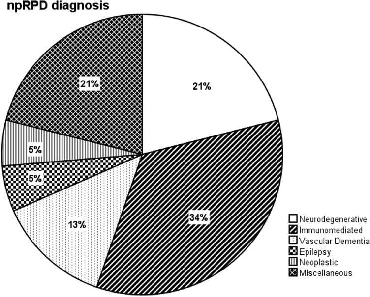
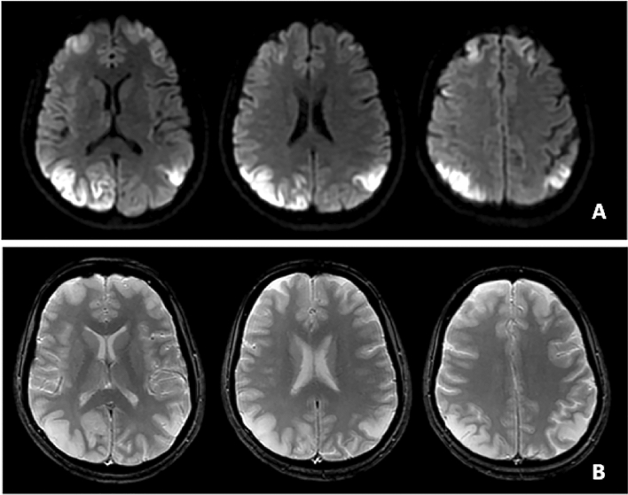
Among patients with definite/probable diagnosis of CJD or genetic prion disease, 2/107 had normal DWI-MRI: in one patient a 2-months follow-up DWI-MRI showed CJD-related changes while the other had autopsy-proven CJD despite no DWI abnormalities 282 days after clinical onset. CJD-related cortical changes were detected in all lobes and involvement of thalamus was common. In the npRPD groups, 6/40 patients showed DWI alterations that clustered in three different patterns: (1) minimal/doubtful signal alterations (limbic encephalitis, dementia with Lewy bodies); (2) clearly suggestive of alternative diagnoses (status epilepticus, Wernicke or metabolic encephalopathy); (3) highly suggestive of CJD (mitochondrial disease), though cortical swelling let exclude CJD.
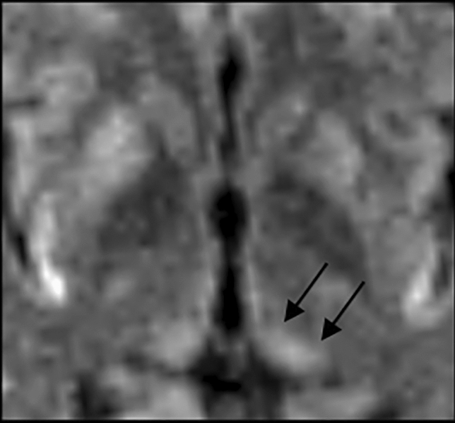
In the diagnostic work-up of RPD, negative/doubtful DWI makes CJD diagnosis rather unlikely, while specific DWI patterns help differentiating CJD from alternative diagnoses. The pulvinar sign is not exclusive of the variant form.
研究伴有和不伴有克雅氏病(CJD)诊断的快速进展性痴呆(RPD)患者队列的脑 MRI 异常。
107 例朊病毒病患者(60 例确诊 sCJD,33 例可能 sCJD,14 例遗传性朊病毒病)和 40 例非朊病毒相关 RPD 患者(npRPD)接受了包括 DWI 和 FLAIR 的脑 MRI 检查。MRI 采用半定量评分进行评估,分别考虑 22 个脑区的异常信号范围和强度。记录了发病时的临床发现、病程、脑脊液 14-3-3 和 t-tau 蛋白水平以及 EEG 数据。
在确诊/可能 CJD 或遗传性朊病毒病患者中,2/107 例 DWI-MRI 正常:1 例患者 2 个月随访 DWI-MRI 显示与 CJD 相关的变化,而另 1 例患者尽管在临床发病后 282 天无 DWI 异常,但尸检证实为 CJD。CJD 相关皮质改变在所有脑叶中均有发现,丘脑受累常见。在 npRPD 组中,6/40 例患者出现 DWI 改变,分为三种不同模式:(1)微小/可疑信号改变(边缘性脑炎,路易体痴呆);(2)明显提示其他诊断(癫痫持续状态,Wernicke 或代谢性脑病);(3)高度提示 CJD(线粒体疾病),但皮质肿胀排除了 CJD。
在 RPD 的诊断工作中,阴性/可疑 DWI 使 CJD 诊断的可能性降低,而特定的 DWI 模式有助于区分 CJD 与其他诊断。丘脑球征并非变异型的特有表现。