Navaneeth G C, Hiremath Rudresh, Poojary Shweta Raviraj, Kini Divya Vishwanatha, Chittaragi Kavitha B
JSS Medical College, Mysuru, Karnataka, India.
Pol J Radiol. 2023 Aug 21;88:e379-e388. doi: 10.5114/pjr.2023.131010. eCollection 2023.
Abdominal obesity plays a significant role in the development of metabolic syndrome, with individual metabolic risk profiles for visceral and subcutaneous adipose tissues. This study aimed to calculate and correlate the subcutaneous, visceral, and total fat compartment volume in metabolic and non-metabolic syndrome patients.
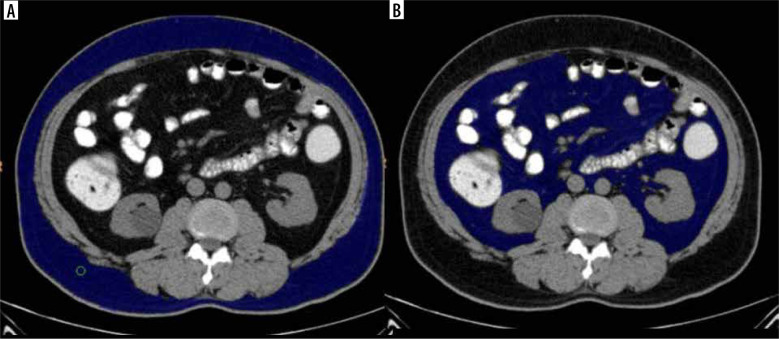
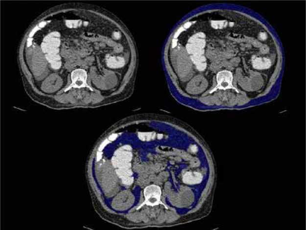
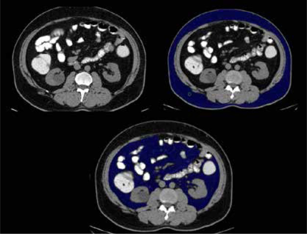
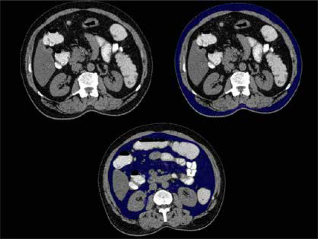
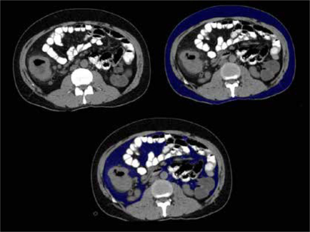
This was a cross-sectional study conducted on 112 patients categorized into Group A (with metabolic syndrome) and Group B (without metabolic syndrome). They were subjected to computed tomography (CT) study of the abdomen using a 128-slice MDCT scanner. Body mass index (BMI), visceral fat volume (VFV), subcutaneous fat volume (SFV), and total fat volume (TFV) were calculated and correlated with biochemical evidence of metabolic syndrome.
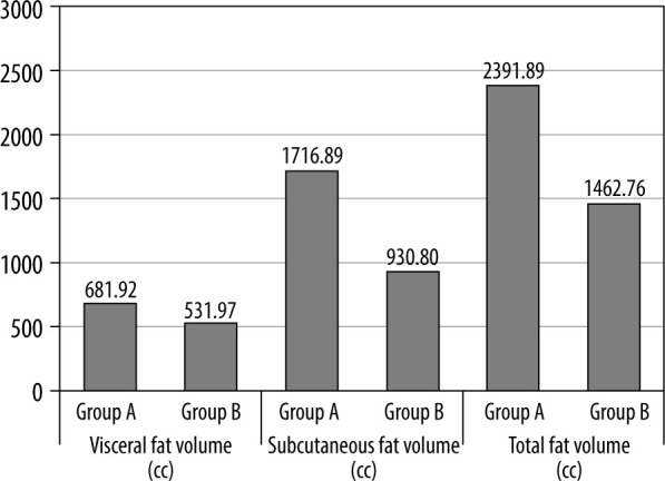
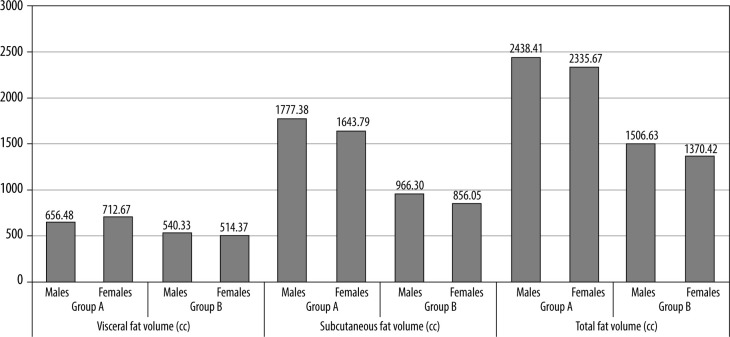
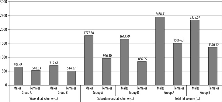
The mean age of patients in Group A was 60.91 ± 12.23 years as compared to Group B, which was 50.12 ± 16.30 years. Overall, a male predominance was observed, i.e. 69 cases (61.6%). BMI was proven to be an inaccurate risk predictor. However, mean VFV, SFV, and TFV was statistically higher in patients with metabolic syndrome ( = 0.001), with visceral fat volume predicting a higher risk in females ( = 0.026).
Abdominal CT is a commonly performed yet unexplored tool for the risk assessment of metabolic syndrome. Through the results obtained in this study, we have proven the need for calculating SFV, VFV, and TFV as predictors of metabolic syndrome in comparison to the conventional practice of BMI assessment. The radiologist can thus work with the clinician to effectively detect and treat this health condition.
腹部肥胖在代谢综合征的发展中起重要作用,内脏和皮下脂肪组织具有个体代谢风险特征。本研究旨在计算代谢综合征患者和非代谢综合征患者的皮下、内脏和总脂肪腔室体积,并进行相关性分析。
这是一项横断面研究,对112例患者进行了分组,分为A组(患有代谢综合征)和B组(未患有代谢综合征)。使用128层MDCT扫描仪对他们进行腹部计算机断层扫描(CT)研究。计算体重指数(BMI)、内脏脂肪体积(VFV)、皮下脂肪体积(SFV)和总脂肪体积(TFV),并与代谢综合征的生化证据进行相关性分析。
A组患者的平均年龄为60.91±12.23岁,而B组为50.12±16.30岁。总体而言,观察到男性占优势,即69例(61.6%)。BMI被证明是一个不准确的风险预测指标。然而,代谢综合征患者的平均VFV、SFV和TFV在统计学上更高(P = 0.001),内脏脂肪体积在女性中预测更高风险(P = 0.026)。
腹部CT是一种常用但尚未充分探索的代谢综合征风险评估工具。通过本研究获得的结果,我们证明了与传统的BMI评估方法相比,计算SFV、VFV和TFV作为代谢综合征预测指标的必要性。放射科医生因此可以与临床医生合作,有效地检测和治疗这种健康状况。