Nagao Itsuma, Ohno Koichi, Nagahara Takuro, Yokoyama Nozomu, Nakagawa Taisuke, Fujiwara Reina, Yamamoto Kie, Goto-Koshino Yuko, Tomiyasu Hirotaka, Tsujimoto Hajime
Department of Veterinary Internal Medicine, Graduate School of Agricultural and Sciences, The University of Tokyo, 1-1-1 Yayoi, Bunkyo-ku, Tokyo 113-0032, Japan.
Veterinary Medical Center, Graduate School of Agricultural and Sciences, The University of Tokyo, 1-1-1 Yayoi, Bunkyo-ku, Tokyo 113-0032, Japan.
J Vet Med Sci. 2019 Nov 14;81(11):1552-1557. doi: 10.1292/jvms.19-0254. Epub 2019 Sep 25.
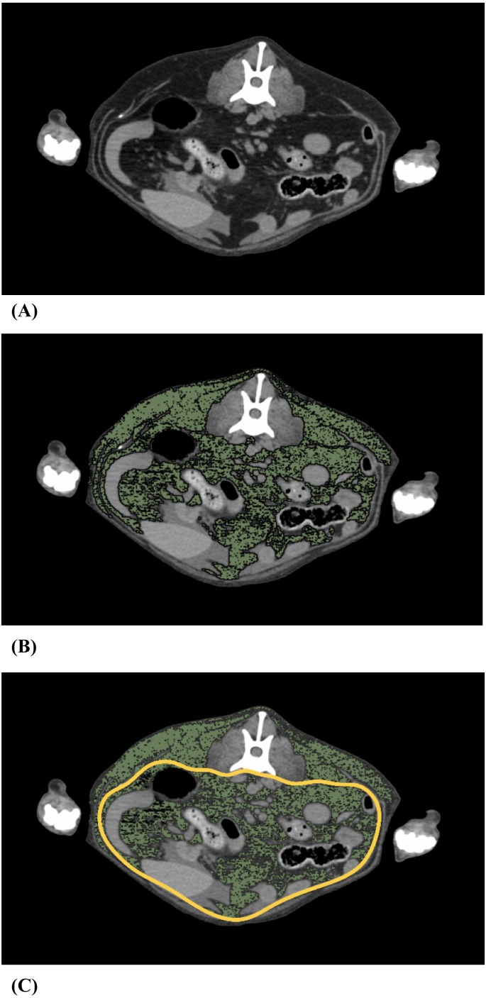
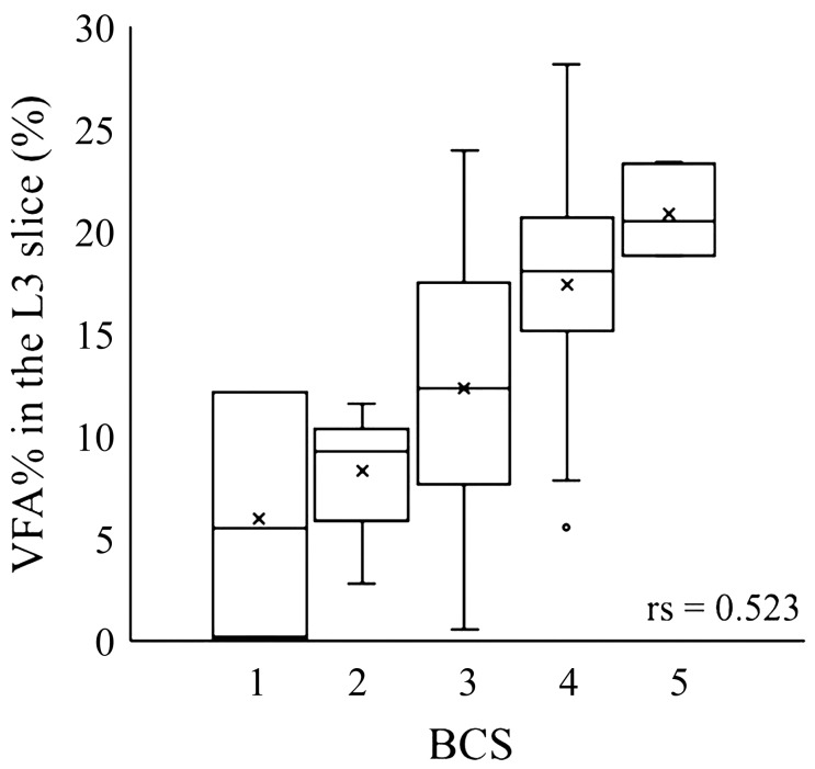
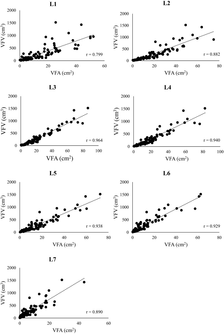
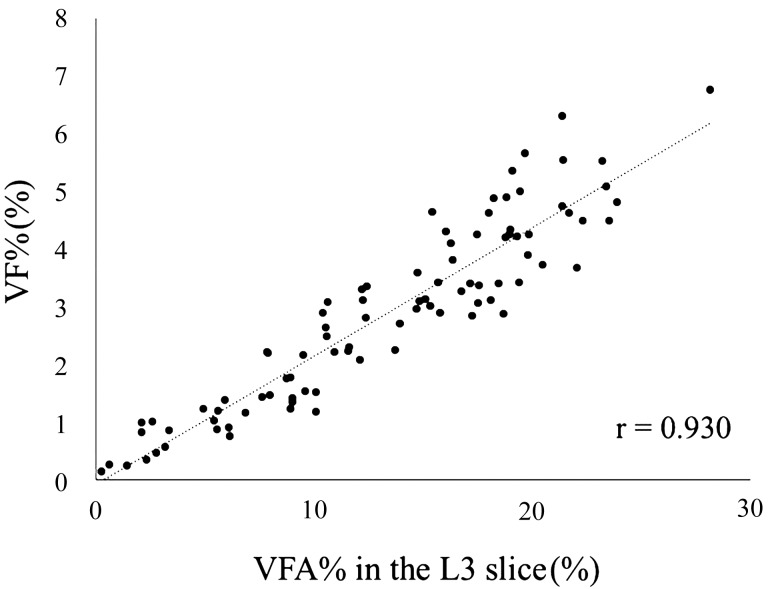
In human medicine, computed tomography (CT) is the gold standard for visceral fat measurement. Research shows that the visceral fat area (VFA) of the umbilical slice is significantly correlated with the visceral fat volume (VFV). In veterinary medicine, however, few studies have evaluated visceral fat using CT. This study aimed to evaluate the visceral fat in dogs using CT images, and determine if the slice significantly correlated with VFV to simplify visceral fat measurements. This retrospective study includes data on 90 dogs that underwent whole-body CT scans for diagnostic purposes. VFV was calculated as the product of VFA and thickness in each CT slice; the correlation between VFV and VFA was analyzed at the level of each lumbar vertebra. Visceral fat percentage (VF%) was calculated as the ratio of the product of VFV and fat density to the body weight. Visceral fat area percentage (VFA%) was calculated as the ratio of VFA to the body area, and its correlation with the VF% and the body condition score (BCS) was analyzed. VFA was highly correlated with VFV at the level of each lumbar vertebra, with the highest correlation (r=0.964) at the L3 level. VFA% was significantly correlated with VF% (r=0.930) and weakly correlated with BCS (r=0.523). This study demonstrates that it is sufficient to use only the L3 slice for visceral fat evaluation and that the evaluation can be based on VFA% of the L3 level.
在人类医学中,计算机断层扫描(CT)是测量内脏脂肪的金标准。研究表明,脐平面的内脏脂肪面积(VFA)与内脏脂肪体积(VFV)显著相关。然而,在兽医学中,很少有研究使用CT评估内脏脂肪。本研究旨在利用CT图像评估犬的内脏脂肪,并确定该平面是否与VFV显著相关,以简化内脏脂肪测量。这项回顾性研究纳入了90只因诊断目的接受全身CT扫描的犬的数据。VFV通过每个CT平面的VFA与厚度的乘积计算得出;在每个腰椎水平分析VFV与VFA之间的相关性。内脏脂肪百分比(VF%)通过VFV与脂肪密度的乘积与体重的比值计算得出。内脏脂肪面积百分比(VFA%)通过VFA与体表面积的比值计算得出,并分析其与VF%和身体状况评分(BCS)的相关性。在每个腰椎水平,VFA与VFV高度相关,在L3水平相关性最高(r=0.964)。VFA%与VF%显著相关(r=0.930),与BCS弱相关(r=0.523)。本研究表明,仅使用L3平面进行内脏脂肪评估就足够了,并且评估可以基于L3水平的VFA%。