• 文献检索
  • 文档翻译
  • 深度研究
  • 学术资讯
  • Suppr Zotero 插件Zotero 插件
  • 邀请有礼
  • 套餐&价格
  • 历史记录
应用&插件
Suppr Zotero 插件Zotero 插件浏览器插件Mac 客户端Windows 客户端微信小程序
定价
高级版会员购买积分包购买API积分包
服务
文献检索文档翻译深度研究API 文档MCP 服务
关于我们
关于 Suppr公司介绍联系我们用户协议隐私条款
关注我们

Suppr 超能文献

核心技术专利:CN118964589B侵权必究
粤ICP备2023148730 号-1Suppr @ 2026

文献检索

告别复杂PubMed语法,用中文像聊天一样搜索,搜遍4000万医学文献。AI智能推荐,让科研检索更轻松。

立即免费搜索

文件翻译

保留排版,准确专业,支持PDF/Word/PPT等文件格式,支持 12+语言互译。

免费翻译文档

深度研究

AI帮你快速写综述,25分钟生成高质量综述,智能提取关键信息,辅助科研写作。

立即免费体验

层流诱导的CHK1-SENP2活性通过不同于内皮-间充质细胞转化的机制阻碍内皮激活和纤维化改变。

Endothelial activation and fibrotic changes are impeded by laminar flow-induced CHK1-SENP2 activity through mechanisms distinct from endothelial-to-mesenchymal cell transition.

作者信息

Nguyen Minh T H, Imanishi Masaki, Li Shengyu, Chau Khanh, Banerjee Priyanka, Velatooru Loka Reddy, Ko Kyung Ae, Samanthapudi Venkata S K, Gi Young J, Lee Ling-Ling, Abe Rei J, McBeath Elena, Deswal Anita, Lin Steven H, Palaskas Nicolas L, Dantzer Robert, Fujiwara Keigi, Borchrdt Mae K, Turcios Estefani Berrios, Olmsted-Davis Elizabeth A, Kotla Sivareddy, Cooke John P, Wang Guangyu, Abe Jun-Ichi, Le Nhat-Tu

机构信息

Center for Cardiovascular Regeneration, Department of Cardiovascular Sciences, Houston Methodist Research Institute, Houston, TX, United States.

Department of Life Science, Vietnam Academy of Science and Technology, University of Science and Technology of Hanoi, Hanoi, Vietnam.

出版信息

Front Cardiovasc Med. 2023 Aug 30;10:1187490. doi: 10.3389/fcvm.2023.1187490. eCollection 2023.

DOI:10.3389/fcvm.2023.1187490
PMID:37711550
原文链接:https://pmc.ncbi.nlm.nih.gov/articles/PMC10499395/
Abstract

BACKGROUND

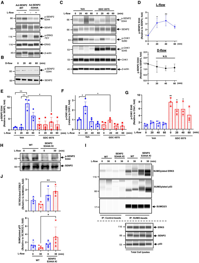
The deSUMOylase sentrin-specific isopeptidase 2 (SENP2) plays a crucial role in atheroprotection. However, the phosphorylation of SENP2 at T368 under disturbed flow (D-flow) conditions hinders its nuclear function and promotes endothelial cell (EC) activation. SUMOylation has been implicated in D-flow-induced endothelial-to-mesenchymal transition (endoMT), but the precise role of SENP2 in counteracting this process remains unclear.

METHOD

We developed a phospho-specific SENP2 S344 antibody and generated knock-in (KI) mice with a phospho-site mutation of SENP2 S344A using CRISPR/Cas9 technology. We then investigated the effects of SENP2 S344 phosphorylation under two distinct flow patterns and during hypercholesteremia (HC)-mediated EC activation.

RESULT

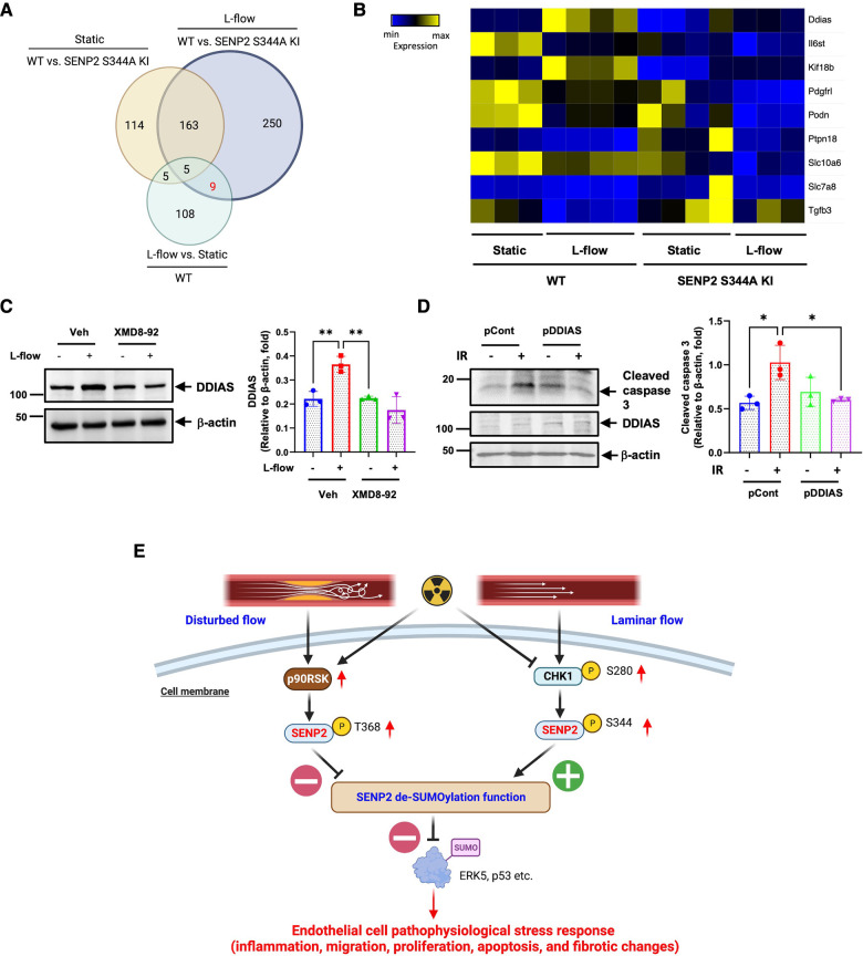
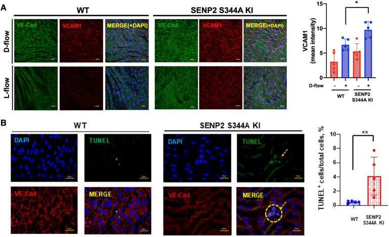
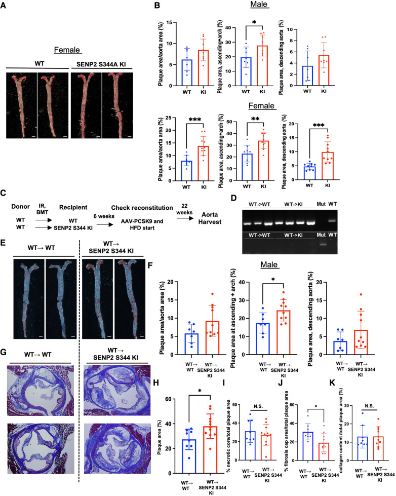
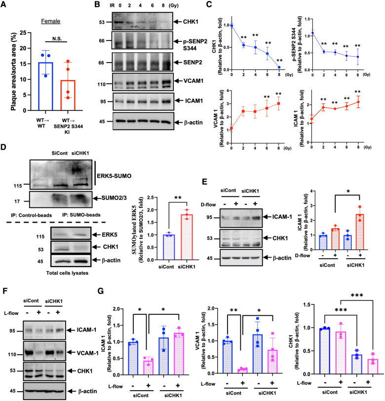
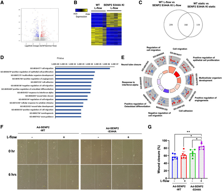
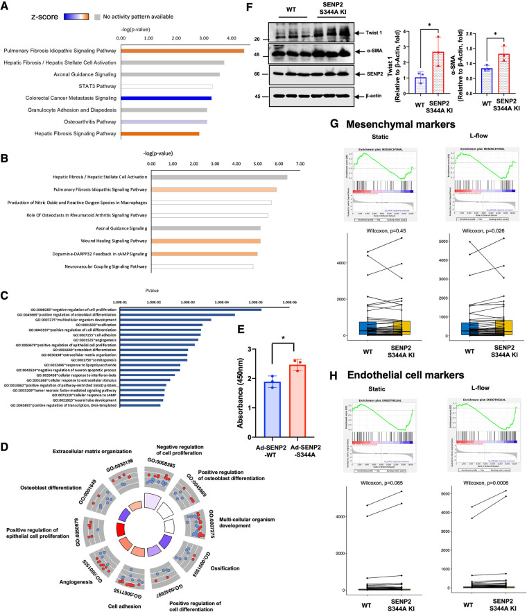
Our findings demonstrate that laminar flow (L-flow) induces phosphorylation of SENP2 at S344 through the activation of checkpoint kinase 1 (CHK1), leading to the inhibition of ERK5 and p53 SUMOylation and subsequent suppression of EC activation. We observed a significant increase in lipid-laden lesions in both the aortic arch (under D-flow) and descending aorta (under L-flow) of female hypercholesterolemic SENP2 S344A KI mice. In male hypercholesterolemic SENP2 S344A KI mice, larger lipid-laden lesions were only observed in the aortic arch area, suggesting a weaker HC-mediated atherogenesis in male mice compared to females. Ionizing radiation (IR) reduced CHK1 expression and SENP2 S344 phosphorylation, attenuating the pro-atherosclerotic effects observed in female SENP2 S344A KI mice after bone marrow transplantation (BMT), particularly in L-flow areas. The phospho-site mutation SENP2 S344A upregulates processes associated with EC activation, including inflammation, migration, and proliferation. Additionally, fibrotic changes and up-regulated expression of EC marker genes were observed. Apoptosis was augmented in ECs derived from the lungs of SENP2 S344A KI mice, primarily through the inhibition of ERK5-mediated expression of DNA damage-induced apoptosis suppressor (DDIAS).

SUMMARY

In this study, we have revealed a novel mechanism underlying the suppressive effects of L-flow on EC inflammation, migration, proliferation, apoptosis, and fibrotic changes through promoting CHK1-induced SENP2 S344 phosphorylation. The phospho-site mutation SENP2 S344A responds to L-flow through a distinct mechanism, which involves the upregulation of both mesenchymal and EC marker genes.

摘要

背景

去SUMO化酶sentrin特异性异肽酶2(SENP2)在动脉粥样硬化保护中起关键作用。然而,在紊乱血流(D-flow)条件下,SENP2第368位苏氨酸(T368)的磷酸化会阻碍其核功能并促进内皮细胞(EC)活化。SUMO化与D-flow诱导的内皮-间充质转化(endoMT)有关,但SENP2在对抗这一过程中的精确作用仍不清楚。

方法

我们开发了一种磷酸化特异性的SENP2 S344抗体,并使用CRISPR/Cas9技术构建了SENP2 S344A磷酸化位点突变的敲入(KI)小鼠。然后,我们研究了在两种不同血流模式下以及高胆固醇血症(HC)介导的EC活化过程中SENP2 S344磷酸化的影响。

结果

我们的研究结果表明,层流(L-flow)通过激活检查点激酶1(CHK1)诱导SENP2在S344位点的磷酸化,导致ERK5和p53 SUMO化的抑制以及随后EC活化的抑制。我们观察到雌性高胆固醇血症SENP2 S344A KI小鼠的主动脉弓(在D-flow下)和降主动脉(在L-flow下)中富含脂质的病变显著增加。在雄性高胆固醇血症SENP2 S344A KI小鼠中,仅在主动脉弓区域观察到更大的富含脂质的病变,这表明与雌性小鼠相比,雄性小鼠中HC介导的动脉粥样硬化发生较弱。电离辐射(IR)降低了CHK1表达和SENP2 S344磷酸化,减轻了雌性SENP2 S344A KI小鼠骨髓移植(BMT)后观察到的促动脉粥样硬化作用,特别是在L-flow区域。磷酸化位点突变SENP2 S344A上调了与EC活化相关的过程,包括炎症、迁移和增殖。此外,还观察到纤维化变化和EC标记基因表达上调。SENP2 S344A KI小鼠肺来源的EC中凋亡增加,主要是通过抑制ERK5介导的DNA损伤诱导凋亡抑制因子(DDIAS)的表达。

总结

在本研究中,我们揭示了一种新机制,即L-flow通过促进CHK1诱导的SENP2 S344磷酸化来抑制EC炎症、迁移、增殖、凋亡和纤维化变化。磷酸化位点突变SENP2 S344A通过一种独特的机制对L-flow作出反应,该机制涉及间充质和EC标记基因的上调。

https://cdn.ncbi.nlm.nih.gov/pmc/blobs/81b7/10499395/1d18ea3e4adb/fcvm-10-1187490-g007.jpg
https://cdn.ncbi.nlm.nih.gov/pmc/blobs/81b7/10499395/f716dc2aec5f/fcvm-10-1187490-g001.jpg
https://cdn.ncbi.nlm.nih.gov/pmc/blobs/81b7/10499395/cf919305f809/fcvm-10-1187490-g002.jpg
https://cdn.ncbi.nlm.nih.gov/pmc/blobs/81b7/10499395/a39efd1b048f/fcvm-10-1187490-g003.jpg
https://cdn.ncbi.nlm.nih.gov/pmc/blobs/81b7/10499395/946316d7f7b8/fcvm-10-1187490-g004.jpg
https://cdn.ncbi.nlm.nih.gov/pmc/blobs/81b7/10499395/b0ec1f22b186/fcvm-10-1187490-g005.jpg
https://cdn.ncbi.nlm.nih.gov/pmc/blobs/81b7/10499395/6578fe9a4fb6/fcvm-10-1187490-g006.jpg
https://cdn.ncbi.nlm.nih.gov/pmc/blobs/81b7/10499395/1d18ea3e4adb/fcvm-10-1187490-g007.jpg

相似文献

1
Endothelial activation and fibrotic changes are impeded by laminar flow-induced CHK1-SENP2 activity through mechanisms distinct from endothelial-to-mesenchymal cell transition.层流诱导的CHK1-SENP2活性通过不同于内皮-间充质细胞转化的机制阻碍内皮激活和纤维化改变。
Front Cardiovasc Med. 2023 Aug 30;10:1187490. doi: 10.3389/fcvm.2023.1187490. eCollection 2023.
2
De-SUMOylation enzyme of sentrin/SUMO-specific protease 2 regulates disturbed flow-induced SUMOylation of ERK5 and p53 that leads to endothelial dysfunction and atherosclerosis.去 SUMO 化酶 SENTRIN/SUMO 特异性蛋白酶 2 调节血流紊乱诱导的 ERK5 和 p53 的 SUMO 化,导致内皮功能障碍和动脉粥样硬化。
Circ Res. 2013 Mar 15;112(6):911-23. doi: 10.1161/CIRCRESAHA.111.300179. Epub 2013 Feb 4.
3
Disturbed flow-activated p90RSK kinase accelerates atherosclerosis by inhibiting SENP2 function.紊乱血流激活的p90RSK激酶通过抑制SENP2功能加速动脉粥样硬化。
J Clin Invest. 2015 Mar 2;125(3):1299-310. doi: 10.1172/JCI76453. Epub 2015 Feb 17.
4
Endothelial Yin Yang 1 Phosphorylation at S118 Induces Atherosclerosis Under Flow.内皮细胞 Yin Yang 1 丝氨酸 118 磷酸化在流动条件下诱导动脉粥样硬化。
Circ Res. 2021 Dec 3;129(12):1158-1174. doi: 10.1161/CIRCRESAHA.121.319296. Epub 2021 Nov 8.
5
Ionizing Radiation Induces Endothelial Inflammation and Apoptosis via p90RSK-Mediated ERK5 S496 Phosphorylation.电离辐射通过p90RSK介导的ERK5 S496磷酸化诱导内皮细胞炎症和凋亡。
Front Cardiovasc Med. 2018 Mar 14;5:23. doi: 10.3389/fcvm.2018.00023. eCollection 2018.
6
MAGI1 as a link between endothelial activation and ER stress drives atherosclerosis.MAGI1 将内皮细胞激活与内质网应激联系起来,从而驱动动脉粥样硬化的发生。
JCI Insight. 2019 Apr 4;4(7). doi: 10.1172/jci.insight.125570.
7
Disturbed Flow-Induced Endothelial Proatherogenic Signaling Via Regulating Post-Translational Modifications and Epigenetic Events.紊乱血流通过调节翻译后修饰和表观遗传事件诱导内皮促动脉粥样硬化信号传导。
Antioxid Redox Signal. 2016 Sep 1;25(7):435-50. doi: 10.1089/ars.2015.6556. Epub 2016 Apr 5.
8
Laminar flow activation of ERK5 leads to cytoprotective effect via CHIP-mediated p53 ubiquitination in endothelial cells.内皮细胞中,ERK5的层流激活通过CHIP介导的p53泛素化产生细胞保护作用。
Anat Cell Biol. 2011 Dec;44(4):265-73. doi: 10.5115/acb.2011.44.4.265. Epub 2011 Dec 30.
9
PKCζ mediates disturbed flow-induced endothelial apoptosis via p53 SUMOylation.PKCζ 通过 p53 SUMOylation 介导紊乱血流诱导的内皮细胞凋亡。
J Cell Biol. 2011 May 30;193(5):867-84. doi: 10.1083/jcb.201010051.
10
An ERK5-NRF2 Axis Mediates Senescence-Associated Stemness and Atherosclerosis.ERK5-NRF2 轴介导衰老相关干性和动脉粥样硬化。
Circ Res. 2023 Jun 23;133(1):25-44. doi: 10.1161/CIRCRESAHA.122.322017. Epub 2023 Jun 2.

引用本文的文献

1
TNIK-driven regulation of ERK5 transcriptional activity in endothelial cells.TNIK驱动的内皮细胞中ERK5转录活性的调控
Front Cardiovasc Med. 2025 Jul 2;12:1526676. doi: 10.3389/fcvm.2025.1526676. eCollection 2025.
2
A visual-omics foundation model to bridge histopathology with spatial transcriptomics.一种将组织病理学与空间转录组学相连接的视觉组学基础模型。
Nat Methods. 2025 May 29. doi: 10.1038/s41592-025-02707-1.
3
A visual-omics foundation model to bridge histopathology image with transcriptomics.一种将组织病理学图像与转录组学相连接的视觉组学基础模型。

本文引用的文献

1
Ionising radiation and cardiovascular disease: systematic review and meta-analysis.电离辐射与心血管疾病:系统综述和荟萃分析。
BMJ. 2023 Mar 8;380:e072924. doi: 10.1136/bmj-2022-072924.
2
Radiation therapy induces immunosenescence mediated by p90RSK.放射治疗会诱导由p90RSK介导的免疫衰老。
Front Cardiovasc Med. 2022 Nov 7;9:988713. doi: 10.3389/fcvm.2022.988713. eCollection 2022.
3
Application of radiation omics in the development of adverse outcome pathway networks: an example of radiation-induced cardiovascular disease.
Res Sq. 2025 Apr 16:rs.3.rs-5183775. doi: 10.21203/rs.3.rs-5183775/v1.
4
Navigating the Maze of Kinases: CaMK-like Family Protein Kinases and Their Role in Atherosclerosis.激酶迷宫导航:钙调蛋白样家族蛋白激酶及其在动脉粥样硬化中的作用。
Int J Mol Sci. 2024 Jun 5;25(11):6213. doi: 10.3390/ijms25116213.
辐射组学在不良结局途径网络开发中的应用:以辐射诱导心血管疾病为例。
Int J Radiat Biol. 2022;98(12):1722-1751. doi: 10.1080/09553002.2022.2110325. Epub 2022 Aug 24.
4
Nucleus-mitochondria positive feedback loop formed by ERK5 S496 phosphorylation-mediated poly (ADP-ribose) polymerase activation provokes persistent pro-inflammatory senescent phenotype and accelerates coronary atherosclerosis after chemo-radiation.ERK5 S496 磷酸化介导的多聚(ADP-核糖)聚合酶激活形成的核-线粒体正反馈环引发持续的促炎衰老表型,并加速化疗-放疗后的冠状动脉粥样硬化。
Redox Biol. 2021 Nov;47:102132. doi: 10.1016/j.redox.2021.102132. Epub 2021 Sep 20.
5
Disturbed flow-induced FAK K152 SUMOylation initiates the formation of pro-inflammation positive feedback loop by inducing reactive oxygen species production in endothelial cells.紊乱流诱导的 FAK K152 SUMOylation 通过诱导内皮细胞产生活性氧,引发促炎正反馈环的形成。
Free Radic Biol Med. 2021 Dec;177:404-418. doi: 10.1016/j.freeradbiomed.2021.09.023. Epub 2021 Oct 5.
6
Endothelial to Mesenchymal Transition: An Insight in Atherosclerosis.内皮细胞向间充质细胞转变:对动脉粥样硬化的深入了解
Front Cardiovasc Med. 2021 Sep 17;8:734550. doi: 10.3389/fcvm.2021.734550. eCollection 2021.
7
Exposure to Low to Moderate Doses of Ionizing Radiation Induces A Reduction of Pro-Inflammatory Ly6chigh Monocytes and a U-Curved Response of T Cells in APOE -/- Mice.暴露于低至中等剂量的电离辐射会导致APOE -/-小鼠中促炎性Ly6c高表达单核细胞减少以及T细胞呈U型反应。
Dose Response. 2021 Jun 8;19(2):15593258211016237. doi: 10.1177/15593258211016237. eCollection 2021 Apr-Jun.
8
The Atr-Chek1 pathway inhibits axon regeneration in response to Piezo-dependent mechanosensation.Atr-Chek1 通路通过抑制 Piezo 依赖性机械感觉来抑制轴突再生。
Nat Commun. 2021 Jun 22;12(1):3845. doi: 10.1038/s41467-021-24131-7.
9
KLF2 Mediates the Suppressive Effect of Laminar Flow on Vascular Calcification by Inhibiting Endothelial BMP/SMAD1/5 Signaling.KLF2 通过抑制内皮细胞 BMP/SMAD1/5 信号通路介导层流对血管钙化的抑制作用。
Circ Res. 2021 Aug 6;129(4):e87-e100. doi: 10.1161/CIRCRESAHA.120.318690. Epub 2021 Jun 23.
10
DNA damage response in vascular endothelial senescence: Implication for radiation-induced cardiovascular diseases.血管内皮衰老中的 DNA 损伤反应:对辐射诱导心血管疾病的影响。
J Radiat Res. 2021 Jul 10;62(4):564-573. doi: 10.1093/jrr/rrab032.