• 文献检索
  • 文档翻译
  • 深度研究
  • 学术资讯
  • Suppr Zotero 插件Zotero 插件
  • 邀请有礼
  • 套餐&价格
  • 历史记录
应用&插件
Suppr Zotero 插件Zotero 插件浏览器插件Mac 客户端Windows 客户端微信小程序
定价
高级版会员购买积分包购买API积分包
服务
文献检索文档翻译深度研究API 文档MCP 服务
关于我们
关于 Suppr公司介绍联系我们用户协议隐私条款
关注我们

Suppr 超能文献

核心技术专利:CN118964589B侵权必究
粤ICP备2023148730 号-1Suppr @ 2026

文献检索

告别复杂PubMed语法,用中文像聊天一样搜索,搜遍4000万医学文献。AI智能推荐,让科研检索更轻松。

立即免费搜索

文件翻译

保留排版,准确专业,支持PDF/Word/PPT等文件格式,支持 12+语言互译。

免费翻译文档

深度研究

AI帮你快速写综述,25分钟生成高质量综述,智能提取关键信息,辅助科研写作。

立即免费体验

使用Sonazoid诊断肝细胞癌:全面综述

Diagnosis of hepatocellular carcinoma using Sonazoid: a comprehensive review.

作者信息

Jeong Woo Kyoung

机构信息

Department of Radiology, Samsung Medical Center, Sungkyunkwan University School of Medicine, Seoul, Korea.

Center for Imaging Sciences, Samsung Medical Center, Sungkyunkwan University School of Medicine, Seoul, Korea.

出版信息

J Liver Cancer. 2023 Sep;23(2):272-283. doi: 10.17998/jlc.2023.08.25. Epub 2023 Sep 19.

DOI:10.17998/jlc.2023.08.25
PMID:37723641
原文链接:https://pmc.ncbi.nlm.nih.gov/articles/PMC10565540/
Abstract

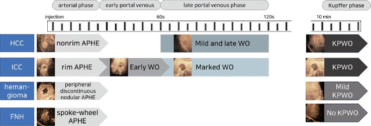
Sonazoid contrast-enhanced ultrasonography (CEUS) is a promising technique for the detection and diagnosis of focal liver lesions, particularly hepatocellular carcinoma (HCC). Recently, a collaborative effort between the Korean Society of Radiology and Korean Society of Abdominal Radiology resulted in the publication of guidelines for diagnosing HCC using Sonazoid CEUS. These guidelines propose specific criteria for identifying HCC based on the imaging characteristics observed during Sonazoid CEUS. The suggested diagnostic criteria include nonrim arterial phase hyperenhancement, and the presence of late and mild washout, or Kupffer phase washout under the premise that the early or marked washout should not occur during the portal venous phase. These criteria aim to improve the accuracy of HCC diagnosis using Sonazoid CEUS. This review offers a comprehensive overview of Sonazoid CEUS in the context of HCC diagnosis. It covers the fundamental principles of Sonazoid CEUS and its clinical applications, and introduces the recently published guidelines. By providing a summary of this emerging technique, this review contributes to a better understanding of the potential role of Sonazoid CEUS for diagnosing HCC.

摘要

舒血管素增强超声造影(CEUS)是一种用于检测和诊断肝脏局灶性病变,尤其是肝细胞癌(HCC)的有前景的技术。最近,韩国放射学会和韩国腹部放射学会共同努力,发表了使用舒血管素CEUS诊断HCC的指南。这些指南根据舒血管素CEUS观察到的影像特征,提出了识别HCC的具体标准。建议的诊断标准包括非边缘动脉期高增强,以及在门静脉期不应出现早期或明显消退的前提下,存在晚期轻度消退或库普弗细胞期消退。这些标准旨在提高使用舒血管素CEUS诊断HCC的准确性。本综述全面概述了舒血管素CEUS在HCC诊断中的应用。它涵盖了舒血管素CEUS的基本原理及其临床应用,并介绍了最近发表的指南。通过总结这种新兴技术,本综述有助于更好地理解舒血管素CEUS在诊断HCC中的潜在作用。

https://cdn.ncbi.nlm.nih.gov/pmc/blobs/4af4/10565540/6d343267c47f/jlc-2023-08-25f7.jpg
https://cdn.ncbi.nlm.nih.gov/pmc/blobs/4af4/10565540/062317ac7bf5/jlc-2023-08-25f1.jpg
https://cdn.ncbi.nlm.nih.gov/pmc/blobs/4af4/10565540/602e66a9a2b2/jlc-2023-08-25f2.jpg
https://cdn.ncbi.nlm.nih.gov/pmc/blobs/4af4/10565540/3b60e66f36b7/jlc-2023-08-25f3.jpg
https://cdn.ncbi.nlm.nih.gov/pmc/blobs/4af4/10565540/20b9f7bb0409/jlc-2023-08-25f4.jpg
https://cdn.ncbi.nlm.nih.gov/pmc/blobs/4af4/10565540/a6fe0b3ddbdb/jlc-2023-08-25f5.jpg
https://cdn.ncbi.nlm.nih.gov/pmc/blobs/4af4/10565540/0a66e8d346c5/jlc-2023-08-25f6.jpg
https://cdn.ncbi.nlm.nih.gov/pmc/blobs/4af4/10565540/6d343267c47f/jlc-2023-08-25f7.jpg

相似文献

1
Diagnosis of hepatocellular carcinoma using Sonazoid: a comprehensive review.使用Sonazoid诊断肝细胞癌:全面综述
J Liver Cancer. 2023 Sep;23(2):272-283. doi: 10.17998/jlc.2023.08.25. Epub 2023 Sep 19.
2
Assessment of arterial-phase hyperenhancement and late-phase washout of hepatocellular carcinoma-a meta-analysis of contrast-enhanced ultrasound (CEUS) with SonoVue® and Sonazoid®.评估肝细胞癌的动脉期增强和晚期廓清: SonoVue® 和 Sonazoid® 超声造影的荟萃分析。
Eur Radiol. 2024 Jun;34(6):3795-3812. doi: 10.1007/s00330-023-10371-2. Epub 2023 Nov 22.
3
Contrast-Enhanced Ultrasound With Perfluorobutane for Hepatocellular Carcinoma Diagnosis: Comparison of Imaging Phases and Diagnostic Criteria.对比增强超声联合全氟丁烷在肝细胞癌诊断中的应用:成像相和诊断标准的比较。
AJR Am J Roentgenol. 2024 Feb;222(2):e2330156. doi: 10.2214/AJR.23.30156. Epub 2023 Nov 22.
4
Impact of Hepatocellular Carcinoma Tumor Size on Sonazoid Contrast-Enhanced Ultrasound Enhancement Features.肝癌肿瘤大小对 SonoVue 超声造影增强特征的影响。
Ultrasound Med Biol. 2024 Jan;50(1):39-46. doi: 10.1016/j.ultrasmedbio.2023.08.022. Epub 2023 Sep 30.
5
The efficacy of modified contrast-enhanced ultrasound Liver Imaging Reporting and Data System (CEUS LI-RADS) using Sonazoid in diagnosis of hepatocellular carcinoma: a systematic review and meta-analysis.使用声诺维的改良对比增强超声肝脏影像报告和数据系统(CEUS LI-RADS)在肝细胞癌诊断中的疗效:一项系统评价和荟萃分析。
Quant Imaging Med Surg. 2024 Apr 3;14(4):2927-2937. doi: 10.21037/qims-23-1184. Epub 2024 Mar 21.
6
Imaging Features of Hepatocellular Carcinoma in the Non-Cirrhotic Liver with Sonazoid-Enhanced Contrast-Enhanced Ultrasound.使用Sonazoid增强型超声造影对非肝硬化肝脏中肝细胞癌的成像特征
Diagnostics (Basel). 2022 Sep 20;12(10):2272. doi: 10.3390/diagnostics12102272.
7
Noninvasive Diagnosis of Hepatocellular Carcinoma on Sonazoid-Enhanced US: Value of the Kupffer Phase.索拉非尼增强超声对肝细胞癌的无创诊断:库普弗细胞期的价值
Diagnostics (Basel). 2022 Jan 7;12(1):141. doi: 10.3390/diagnostics12010141.
8
Diagnosing Hepatocellular Carcinoma Using Sonazoid Contrast-Enhanced Ultrasonography: 2023 Guidelines From the Korean Society of Radiology and the Korean Society of Abdominal Radiology.使用 SonoVue 对比增强超声诊断肝细胞癌:韩国放射学会和韩国腹部放射学会 2023 年指南。
Korean J Radiol. 2023 Jun;24(6):482-497. doi: 10.3348/kjr.2023.0324.
9
Sonazoid-enhanced ultrasonography: comparison with CT/MRI Liver Imaging Reporting and Data System in patients with suspected hepatocellular carcinoma.声诺维增强超声检查:与疑似肝细胞癌患者的CT/MRI肝脏影像报告和数据系统的比较
Ultrasonography. 2021 Oct;40(4):486-498. doi: 10.14366/usg.20120. Epub 2021 Jan 15.
10
B-Mode Ultrasonography versus Contrast-Enhanced Ultrasonography for Surveillance of Hepatocellular Carcinoma: A Prospective Multicenter Randomized Controlled Trial.B超与超声造影在肝细胞癌监测中的对比:一项前瞻性多中心随机对照试验
Liver Cancer. 2019 Jul;8(4):271-280. doi: 10.1159/000501082. Epub 2019 Jun 6.

引用本文的文献

1
Echo contrast medium: How the use of contrast echocardiography (ultrasound contrast agents) can improve patient care.超声造影剂:超声心动图造影(超声造影剂)的应用如何改善患者护理。
World J Methodol. 2025 Sep 20;15(3):100490. doi: 10.5662/wjm.v15.i3.100490.
2
Differentiation of AFP-negative hepatocellular carcinoma from other intrahepatic malignant lesions by a noninvasive predictive model based on Sonazoid contrast-enhanced ultrasound.基于Sonazoid对比增强超声的非侵入性预测模型对甲胎蛋白阴性肝细胞癌与其他肝内恶性病变的鉴别诊断
Front Oncol. 2025 Jul 17;15:1623670. doi: 10.3389/fonc.2025.1623670. eCollection 2025.
3

本文引用的文献

1
2022 KLCA-NCC Korea practice guidelines for the management of hepatocellular carcinoma.《2022年韩国肝脏癌协会-韩国国立癌症中心肝细胞癌管理实践指南》
J Liver Cancer. 2023 Mar;23(1):1-120. doi: 10.17998/jlc.2022.11.07. Epub 2022 Dec 9.
2
Diagnosing Hepatocellular Carcinoma Using Sonazoid Contrast-Enhanced Ultrasonography: 2023 Guidelines From the Korean Society of Radiology and the Korean Society of Abdominal Radiology.使用 SonoVue 对比增强超声诊断肝细胞癌:韩国放射学会和韩国腹部放射学会 2023 年指南。
Korean J Radiol. 2023 Jun;24(6):482-497. doi: 10.3348/kjr.2023.0324.
3
AASLD Practice Guidance on prevention, diagnosis, and treatment of hepatocellular carcinoma.
The Diagnostic Impact of Contrast-Enhanced Computed Tomography (CECT) in Evaluating Lymph Node Involvement in Colorectal Cancer: A Comprehensive Review.
对比增强计算机断层扫描(CECT)在评估结直肠癌淋巴结受累中的诊断价值:一项综述
Cureus. 2024 Jun 6;16(6):e61832. doi: 10.7759/cureus.61832. eCollection 2024 Jun.
4
Sonazoid contrast-enhanced ultrasonography for the diagnosis of hepatocellular carcinoma: strengths and shortcomings.用于诊断肝细胞癌的声诺维增强超声检查:优势与不足
J Liver Cancer. 2023 Sep;23(2):238-240. doi: 10.17998/jlc.2023.09.12. Epub 2023 Sep 20.
美国肝病研究学会肝细胞癌预防、诊断和治疗实践指南。
Hepatology. 2023 Dec 1;78(6):1922-1965. doi: 10.1097/HEP.0000000000000466. Epub 2023 May 22.
4
2022 KLCA-NCC Korea Practice Guidelines for the Management of Hepatocellular Carcinoma.2022KLCA-NCC 韩国肝细胞癌管理实践指南。
Korean J Radiol. 2022 Dec;23(12):1126-1240. doi: 10.3348/kjr.2022.0822.
5
Perfluorobutane-enhanced ultrasonography with a Kupffer phase: improved diagnostic sensitivity for hepatocellular carcinoma.含氟丁烷增强超声造影的 Kupffer 期成像:提高肝细胞癌的诊断灵敏度。
Eur Radiol. 2022 Dec;32(12):8507-8517. doi: 10.1007/s00330-022-08900-6. Epub 2022 Jun 16.
6
Diagnostic criteria of perfluorobutane-enhanced ultrasonography for diagnosing hepatocellular carcinoma in high-risk individuals: how is late washout determined?全氟丁烷增强超声检查诊断高危人群肝细胞癌的诊断标准:如何确定延迟洗脱?
Ultrasonography. 2022 Jul;41(3):530-542. doi: 10.14366/usg.21172. Epub 2021 Dec 17.
7
How to Use LI-RADS to Report Liver CT and MRI Observations.LI-RADS 用于报告肝脏 CT 和 MRI 观察结果的方法。
Radiographics. 2021 Sep-Oct;41(5):1352-1367. doi: 10.1148/rg.2021200205. Epub 2021 Jul 23.
8
Use of Contrast-Enhanced Ultrasound with Sonazoid for Evaluating the Radiotherapy Efficacy for Hepatocellular Carcinoma.使用含声诺维的超声造影评估肝细胞癌的放射治疗疗效
Diagnostics (Basel). 2021 Mar 9;11(3):486. doi: 10.3390/diagnostics11030486.
9
Sonazoid-enhanced ultrasonography: comparison with CT/MRI Liver Imaging Reporting and Data System in patients with suspected hepatocellular carcinoma.声诺维增强超声检查:与疑似肝细胞癌患者的CT/MRI肝脏影像报告和数据系统的比较
Ultrasonography. 2021 Oct;40(4):486-498. doi: 10.14366/usg.20120. Epub 2021 Jan 15.
10
A study on the inconsistency of arterial phase hypervascularity detection between contrast-enhanced ultrasound using sonazoid and gadolinium-ethoxybenzyl-diethylenetriamine penta-acetic acid magnetic resonance imaging of hepatocellular carcinoma lesions.一项关于使用 SonoVue 超声造影与钆塞酸二葡甲胺磁共振成像检测肝细胞癌病灶动脉期高增强一致性的研究。
J Med Ultrason (2001). 2021 Apr;48(2):215-224. doi: 10.1007/s10396-021-01086-2. Epub 2021 Mar 15.