Department of Endodontics, Nanjing Stomatological Hospital, Affiliated Hospital of Medical School, Nanjing University, Nanjing, 210008, Jiangsu, People's Republic of China.
Nanjing Tongren Hospital, Nanjing, 210008, Jiangsu, People's Republic of China.
Sci Rep. 2023 Sep 19;13(1):15512. doi: 10.1038/s41598-023-42767-x.
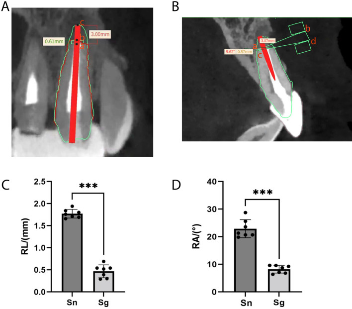
We aimed to design a novel three-dimensional (3D) printed surgical guide and evaluate its accuracy in assisting endodontic microsurgeries. A new 3D printed surgical guide was designed by computer-aided design and computer-aided manufacturing (CAD/CAM) technology and applied to 7 patients who underwent endodontic microsurgeries of upper anterior teeth from 2020.01 to 2020.12 as the experimental group. 7 patients who suffered from endodontic microsurgeries operated by the same surgeon without using the surgical guide from 2019.01 to 2019.12 were selected as the control group. Cone beam computed tomography (CBCT) was performed more than 12 months after operation, and the accuracy of apical resection was compared between the two groups. The accuracy of the microsurgery focused on the length and angle of the root apical resection. In the study, CBCT data and oral digital scanning data were used to reconstruct 3D models of periapical lesions with soft and hard tissue information, based on which we designed the new 3D printed surgical guides. The guides were successfully applied to the apectomy in endodontic microsurgeries. The deviation of the apical resection length of the experimental group (0.467 ± 0.146 mm) was better than that of the control group (1.743 ± 0.122 mm) (P < 0.0001), and the deviation of the apical resection angle of the experimental group (9.711 ± 3.593°) was significantly less than that of the control group (22.400 ± 3.362°) (P < 0.0001). The 3D-printed surgical guide could effectively guide endodontic microsurgery and improve its accuracy by fixing both the position and the angle of apectomy. The new type of surgical guide could accurately localize the root apex and guide the apical resection.
我们旨在设计一种新型的三维(3D)打印手术导板,并评估其在辅助牙髓微创手术中的准确性。通过计算机辅助设计和计算机辅助制造(CAD/CAM)技术设计了一种新的 3D 打印手术导板,并将其应用于 2020 年 1 月至 2020 年 12 月期间接受上前牙牙髓微创手术的 7 例患者(实验组)。选择 2019 年 1 月至 2019 年 12 月期间由同一位外科医生进行手术但未使用手术导板的 7 例患者作为对照组。术后超过 12 个月进行锥形束 CT(CBCT)检查,比较两组根尖切除的准确性。手术精度主要集中在根尖切除的长度和角度。在本研究中,使用 CBCT 数据和口腔数字扫描数据,基于软组织和硬组织信息重建了根尖病变的 3D 模型,在此基础上设计了新的 3D 打印手术导板。导板成功应用于牙髓微创手术中的根尖切除术。实验组根尖切除长度的偏差(0.467 ± 0.146 mm)优于对照组(1.743 ± 0.122 mm)(P < 0.0001),实验组根尖切除角度的偏差(9.711 ± 3.593°)明显小于对照组(22.400 ± 3.362°)(P < 0.0001)。3D 打印手术导板可以通过固定根尖切除的位置和角度,有效指导牙髓微创手术,提高手术精度。新型手术导板能够精确定位根尖,并引导根尖切除。