Cen Chunyuan, Wang Chunyou, Wang Siqi, Wen Kan, Liu Liying, Li Xin, Wu Linxia, Huang Mengting, Ma Ling, Liu Huan, Wu Heshui, Han Ping
Department of Radiology, Union Hospital, Tongji Medical College, Huazhong University of Science and Technology, Wuhan, Hubei, China.
Hubei Province Key Laboratory of Molecular Imaging, Wuhan, Hubei, China.
Front Oncol. 2023 Sep 4;13:1218128. doi: 10.3389/fonc.2023.1218128. eCollection 2023.
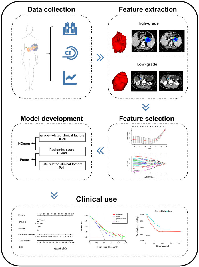
Tumor grading is important for prognosis of pancreatic ductal adenocarcinoma (PDAC). In this study, we developed preoperative clinical-radiomics nomograms using features from contrast-enhanced CT (CECT), to discriminate high-grade and low-grade PDAC and predict overall survival (OS).
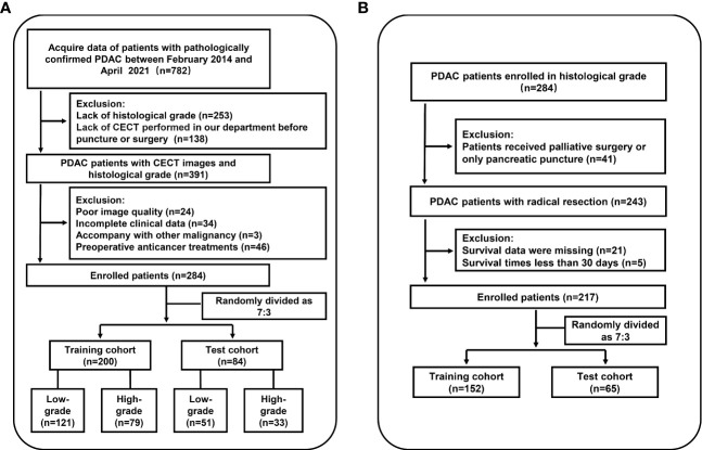
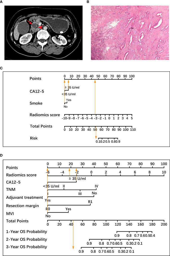
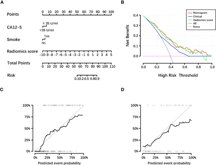
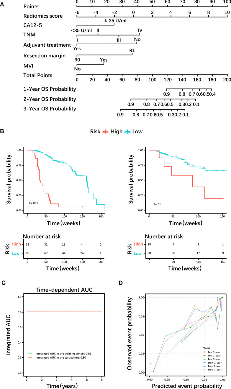
In this single-center, retrospective study conducted from February 2014 to April 2021, consecutive PDAC patients who underwent CECT and had pathologically identified grading were randomized to training (n=200) and test (n=84) cohorts for development of model to predict histological grade based on radiomics scores from CECT (HGrad). Another 42 patients were used as external validation cohort of HGrad. A nomogram (HGnom) was constructed using radiomics score, CA12-5 and smoking to predict histological grade. A second nomogram (Pnom) was constructed using radiomics score, CA12-5, TNM, adjuvant treatment, resection margin and microvascular invasion to predict OS in radical resection patients (217 of 284).
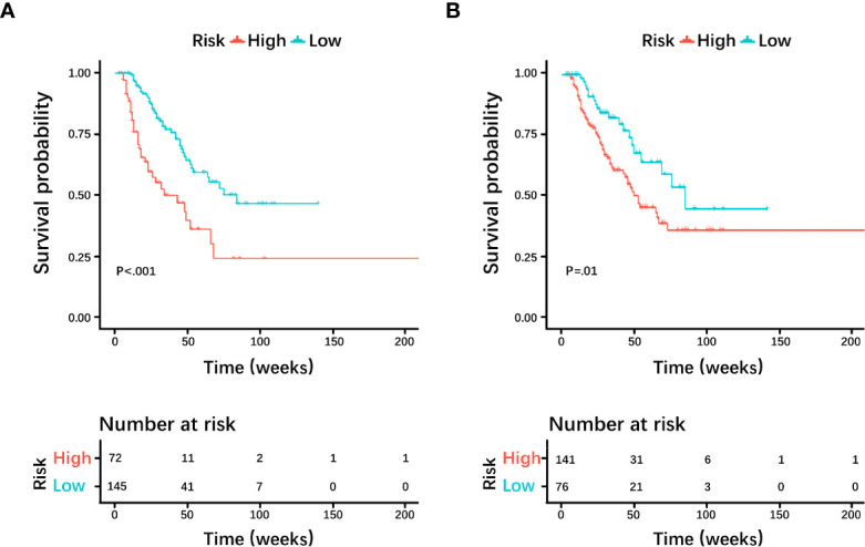
Among 326 patients, 122 were high-grade (120 poorly differentiated and 2 undifferentiated). The HGrad yielded AUCs of 0.75 (95% CI: 0.64, 0.85) and 0.76 (95% CI: 0.60, 0.91) in test and validation cohorts. The HGnom achieved AUCs of 0.77 (95% CI: 0.66, 0.87), and the predicted grades calibrated well with actual grades (=.13). OS was different between the grades predicted by radiomics scores (=.01). The integrated AUC of the Pnom for predicting OS was 0.80 (95% CI: 0.75, 0.88).
Compared with the HGrad using features from CECT, the HGnom demonstrated higher performance for predicting histological grade. The Pnom helped identify patients with high survival outcome in pancreatic ductal adenocarcinoma.
肿瘤分级对胰腺导管腺癌(PDAC)的预后很重要。在本研究中,我们利用对比增强CT(CECT)的特征开发了术前临床-影像组学列线图,以区分高级别和低级别PDAC并预测总生存期(OS)。
在这项于2014年2月至2021年4月进行的单中心回顾性研究中,将接受CECT检查且病理分级明确的连续PDAC患者随机分为训练组(n = 200)和测试组(n = 84),用于开发基于CECT影像组学评分(HGrad)预测组织学分级的模型。另外42例患者用作HGrad的外部验证队列。使用影像组学评分、CA12-5和吸烟情况构建列线图(HGnom)来预测组织学分级。使用影像组学评分、CA12-5、TNM、辅助治疗、切缘和微血管侵犯构建第二个列线图(Pnom)来预测根治性切除患者(284例中的217例)的OS。
在326例患者中,122例为高级别(120例低分化和2例未分化)。HGrad在测试组和验证组中的AUC分别为0.75(95%CI:0.64,0.85)和0.76(95%CI:0.60,0.91)。HGnom的AUC为0.77(95%CI:0.66,0.87),预测分级与实际分级校准良好(P =.13)。影像组学评分预测的不同分级之间OS存在差异(P =.01)。Pnom预测OS的综合AUC为0.80(95%CI:0.75,0.88)。
与使用CECT特征的HGrad相比,HGnom在预测组织学分级方面表现出更高的性能。Pnom有助于识别胰腺导管腺癌中生存结局良好的患者。