Singh Balbir, Wang Zhengyang, Madiah Leen M, Gatti S Elizabeth, Fulton Jenna N, Johnson Graham W, Li Rui, Dawant Benoit M, Englot Dario J, Bick Sarah K, Roberson Shawniqua Williams, Constantinidis Christos
Department of Biomedical Engineering, Vanderbilt University.
Neuroscience Program, Vanderbilt University.
bioRxiv. 2023 Sep 7:2023.09.06.556554. doi: 10.1101/2023.09.06.556554.
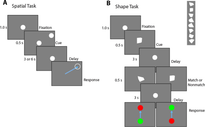
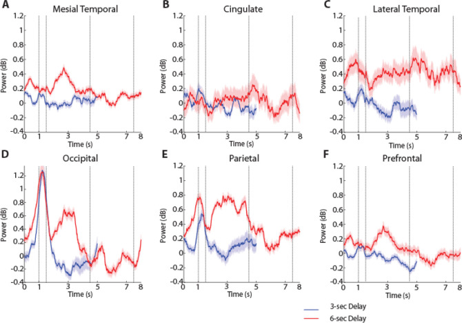
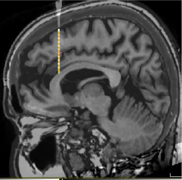
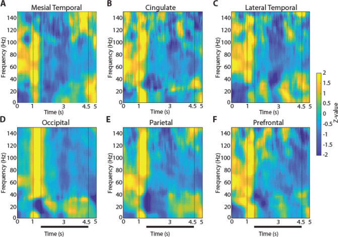
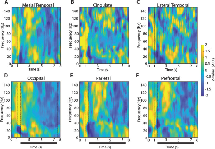
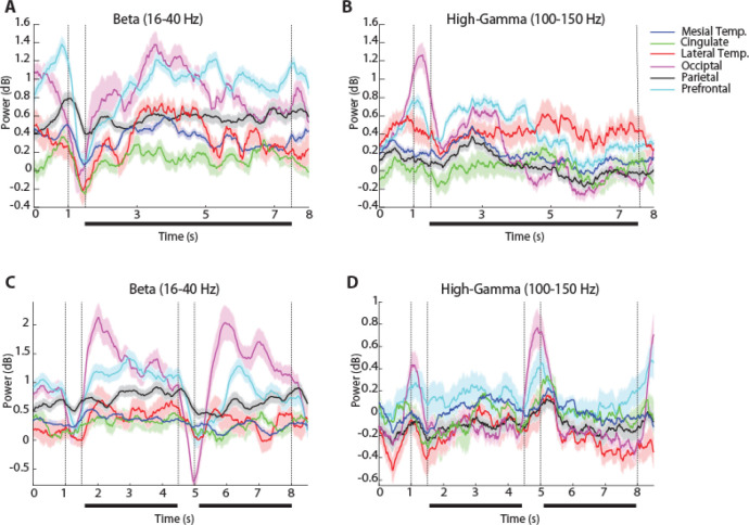
Oscillatory activity is thought to be a marker of cognitive processes, although its role and distribution across the brain during working memory has been a matter of debate. To understand how oscillatory activity differentiates tasks and brain areas in humans, we recorded local field potentials (LFPs) in 12 adults as they performed visual-spatial and shape-matching memory tasks. Tasks were designed to engage working memory processes at a range of delay intervals between stimulus delivery and response initiation. LFPs were recorded using intracranial depth electrodes implanted to localize seizures for management of intractable epilepsy. Task-related LFP power analyses revealed an extensive network of cortical regions that were activated during the presentation of visual stimuli and during their maintenance in working memory, including occipital, parietal, temporal, insular, and prefrontal cortical areas, and subcortical structures including the amygdala and hippocampus. Across most brain areas, the appearance of a stimulus produced broadband power increase, while gamma power was evident during the delay interval of the working memory task. Notable differences between areas included that occipital cortex was characterized by elevated power in the high gamma (100-150 Hz) range during the 500 ms of visual stimulus presentation, which was less pronounced or absent in other areas. A decrease in power centered in beta frequency (16-40 Hz) was also observed after the stimulus presentation, whose magnitude differed across areas. These results reveal the interplay of oscillatory activity across a broad network, and region-specific signatures of oscillatory processes associated with visual working memory.
振荡活动被认为是认知过程的一个标志,尽管其在工作记忆期间在大脑中的作用和分布一直存在争议。为了了解振荡活动如何区分人类的任务和脑区,我们记录了12名成年人在执行视觉空间和形状匹配记忆任务时的局部场电位(LFP)。这些任务旨在在刺激呈现和反应启动之间的一系列延迟间隔内激发工作记忆过程。使用植入的颅内深度电极记录LFP,以定位癫痫发作,用于治疗难治性癫痫。与任务相关的LFP功率分析揭示了一个广泛的皮质区域网络,这些区域在视觉刺激呈现期间和在工作记忆中维持刺激时被激活,包括枕叶、顶叶、颞叶、岛叶和前额叶皮质区域,以及包括杏仁核和海马体在内的皮质下结构。在大多数脑区,刺激的出现会导致宽带功率增加,而在工作记忆任务的延迟间隔期间,伽马功率很明显。各区域之间的显著差异包括,枕叶皮质在视觉刺激呈现的500毫秒期间,在高伽马(100 - 150赫兹)范围内具有升高的功率,这在其他区域不太明显或不存在。在刺激呈现后,还观察到以β频率(16 - 40赫兹)为中心的功率下降,其幅度因区域而异。这些结果揭示了广泛网络中振荡活动的相互作用,以及与视觉工作记忆相关的振荡过程的区域特异性特征。