Takahashi Tomonari, Igarashi-Yokoi Tae, Nakao Noriko, Uramoto Kengo, Yoshida Takeshi, Ohno-Matsui Kyoko
Department of Ophthalmology and Visual Science, Tokyo Medical and Dental University, 1-5-45 Yushima, Bunkyo-ku, Tokyo, 113-8510, Japan.
Am J Ophthalmol Case Rep. 2023 Sep 4;32:101926. doi: 10.1016/j.ajoc.2023.101926. eCollection 2023 Dec.
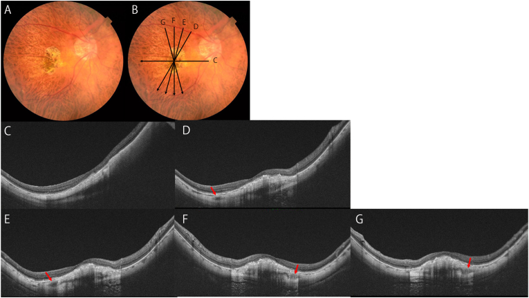
To report our findings in a patient with a two layered dome shaped macula (DSM) in which only the inner layer of the sclera protruded anteriorly.
An 84-year-old woman with high myopia had a DSM in both eyes. The optical coherence tomographic (OCT) image of the left eye showed a uniform thickening of the foveal sclera, but the DSM of the right eye was split into an inner and outer layer by intrascleral blood vessels running between the two layers. OCT showed that only the inner layer of the sclera protruded anteriorly while the outer layer remained in its normal position.
The two layered DSM suggests that the etiology of DSMs may be more complex.
报告我们在一名患有双层圆顶状黄斑(DSM)患者中的发现,该患者仅巩膜内层向前突出。
一名患有高度近视的84岁女性双眼均有DSM。左眼的光学相干断层扫描(OCT)图像显示黄斑区巩膜均匀增厚,但右眼的DSM被两层之间穿行的巩膜内血管分为内层和外层。OCT显示仅巩膜内层向前突出,而外层保持在其正常位置。
双层DSM提示DSM的病因可能更为复杂。