Dan Alexandra Oltea, Bălășoiu Andrei Theodor, Puiu Ileana, Tănasie Andreea Cornelia, Târtea Anca Elena, Sfredel Veronica
Department of Physiology, University of Medicine and Pharmacy of Craiova, 200349 Craiova, Romania.
Department of Ophthalmology, University of Medicine and Pharmacy of Craiova, 200349 Craiova, Romania.
Diagnostics (Basel). 2023 Sep 13;13(18):2934. doi: 10.3390/diagnostics13182934.
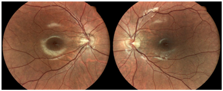
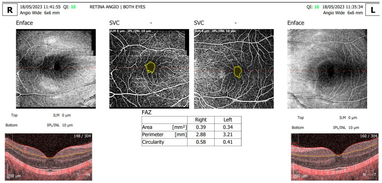
Type 1 diabetes mellitus (type 1 DM) is one of the most prevalent endocrinological diseases among children and young adults, with a growing incidence rate reaching up to 2.9 new cases per year per 100,000 persons below 15 years of age. We report a rare case of a 20-year-old female patient with type 1 DM, hemoglobin D (HbD) heterozygote variant and high myopia of -10.00 spheric diopters, and describe the retinal microvascular alterations visible on OCT angiography (angio-OCT). The patient also presented with a severe stature deficit (less than three standard deviations) and delayed puberty, which could not be explained only by suboptimal glycemic control and indicated possible hypopituitarism. HbA1c level evaluated with the high-performance liquid chromatography (HPLC) method was 6.5%, a falsely low value due to HbD hemoglobinopathy. On ophthalmic evaluation, the angio-OCT scan showed the following retinal microvascular alterations in the right eye (RE): the FAZ (Foveal Avascular Zone) area was 0.39 mm, the FAZ perimeter was 2.88 mm, and the circularity index was 0.58. The following alterations were shown in the left eye (LE): the FAZ area was 0.34 mm, the FAZ perimeter was 3.21 mm, and the circularity index was 0.41. Clinicians should consider high-performance retinal screening methods such as angio-OCT evaluation for young type 1 DM patients, especially for those with associated pathologies like high myopia and hemoglobinopathies. Moreover, multiple evaluation methods of HbA1c values are mandatory as hemoglobinopathies can interfere with the accuracy of HbA1c assay methods.
1型糖尿病(T1DM)是儿童和青年中最常见的内分泌疾病之一,发病率不断上升,在15岁以下人群中每年每10万人高达2.9例新发病例。我们报告了一例罕见病例,一名20岁女性T1DM患者,血红蛋白D(HbD)杂合子变异,近视高达-10.00球镜,并描述了光学相干断层扫描血管造影(angio-OCT)可见的视网膜微血管改变。该患者还存在严重的身材矮小(低于三个标准差)和青春期延迟,这不能仅用血糖控制不佳来解释,提示可能存在垂体功能减退。用高效液相色谱(HPLC)法评估的糖化血红蛋白(HbA1c)水平为6.5%,由于HbD血红蛋白病,该值为假性低值。眼科评估时,angio-OCT扫描显示右眼(RE)有以下视网膜微血管改变:黄斑无血管区(FAZ)面积为0.39mm,FAZ周长为2.88mm,圆度指数为0.58。左眼(LE)有以下改变:FAZ面积为0.34mm,FAZ周长为3.21mm,圆度指数为0.41。临床医生应考虑对年轻的T1DM患者采用如angio-OCT评估等高效的视网膜筛查方法,特别是对于那些伴有高度近视和血红蛋白病等相关病变的患者。此外,由于血红蛋白病会干扰HbA1c检测方法的准确性,因此必须采用多种HbA1c值评估方法。