Pérez Inmaculada, Galindo Sara, López-Miguel Alberto, Nieto-Miguel Teresa, de la Mata Ana, López-Paniagua Marina, Alberca Mercedes, Herreras José M, Calonge Margarita
IOBA (Institute of Applied Ophthalmobiology), Universidad de Valladolid, Campus Miguel Delibes, Paseo Belén, 17, 47011, Valladolid, Spain.
CIBER-BBN (Biomedical Research Networking Center in Bioengineering, Biomaterials and Nanomedicine), Carlos III National Institute of Health, Valladolid, Spain.
Ophthalmol Ther. 2023 Dec;12(6):3251-3262. doi: 10.1007/s40123-023-00809-7. Epub 2023 Sep 29.
The aim of this work is to evaluate the effect of mesenchymal stem cell transplantation (MSCT) and cultivated limbal epithelial transplantation (CLET) therapies on the limbus of patients suffering from limbal stem cell deficiency (LSCD).
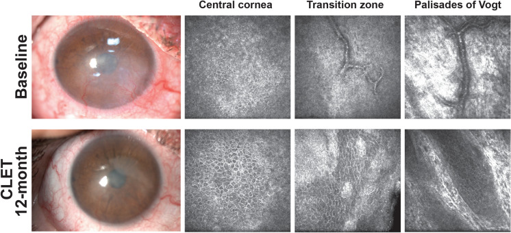
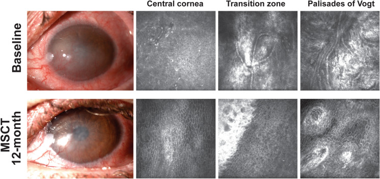
A sub-analysis of a phase I-II randomized, controlled, and double-masked clinical trial was performed to assess the changes in the anatomical structures of the limbus. In vivo confocal microscopy (IVCM) analysis was carried out in LSCD eyes before and 12 months after allogeneic MSCT or CLET. Epithelial phenotype of the central cornea, as well as the presence of transition zones and palisades of Vogt in the limbus, were assessed using Wilcoxon test.
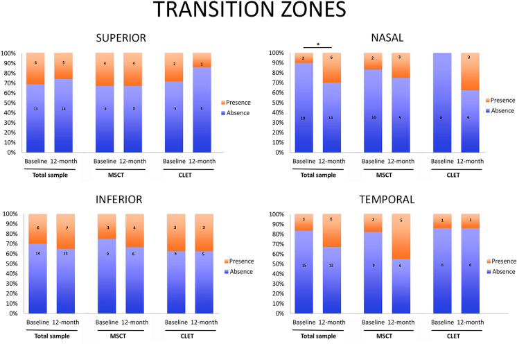
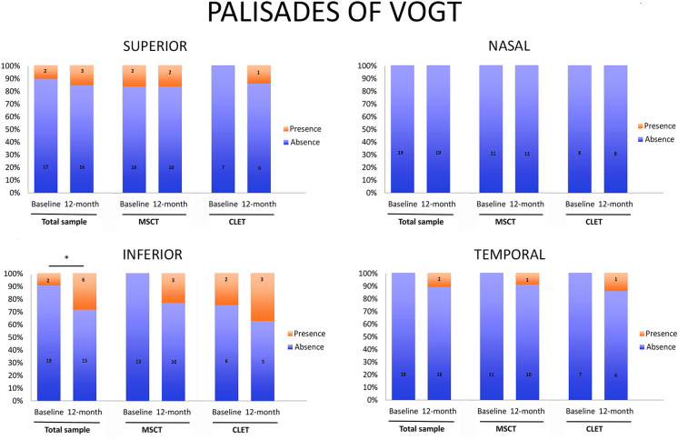
Twenty-three LSCD (14 MSCT and nine CLET) eyes were included. The epithelial phenotype of the central cornea improved significantly (p < 0.001) from 15 (eight MSCT, seven CLET) and eight (six MSCT, two CLET) LSCD eyes showing conjunctival and mixed phenotypes, respectively, to eight (five MSCT, three CLET), five (two MSCT, three CLET), and ten (seven MSCT, three CLET) eyes showing conjunctival, mixed, and corneal phenotypes, respectively. Transition areas and palisades of Vogt were observed in at least one quadrant in nine (five MSCT, four CLET) and 16 (nine MSCT, seven CLET), and in four (two MSCT, two CLET) and six (three MSCT, three CLET) LSCD eyes before and after surgery, respectively. Changes in the transition zones and palisades were solely significant (p = 0.046) for the nasal and inferior quadrants, respectively.
MSCT and CLET improved the central corneal epithelial phenotype despite only minor changes in the anatomical structures of the limbus, as detected by IVCM technology.
ClinicalTrials.gov identifier, NCT01562002.
本研究旨在评估间充质干细胞移植(MSCT)和培养的角膜缘上皮移植(CLET)疗法对角膜缘干细胞缺乏(LSCD)患者角膜缘的影响。
对一项I-II期随机、对照、双盲临床试验进行亚分析,以评估角膜缘解剖结构的变化。在同种异体MSCT或CLET治疗前及治疗后12个月,对LSCD患者的眼睛进行活体共聚焦显微镜(IVCM)分析。使用Wilcoxon检验评估中央角膜的上皮表型,以及角膜缘过渡区和Vogt栅栏的存在情况。
纳入23只LSCD眼睛(14只接受MSCT,9只接受CLET)。中央角膜的上皮表型有显著改善(p < 0.001),分别有15只(8只接受MSCT,7只接受CLET)和8只(6只接受MSCT,2只接受CLET)LSCD眼睛术前表现为结膜和混合表型,术后分别变为8只(5只接受MSCT,3只接受CLET)、5只(2只接受MSCT,3只接受CLET)和10只(7只接受MSCT,3只接受CLET)眼睛,分别表现为结膜、混合和角膜表型。术前和术后分别有9只(5只接受MSCT,4只接受CLET)和16只(9只接受MSCT,7只接受CLET)LSCD眼睛在至少一个象限观察到过渡区和Vogt栅栏,术前和术后分别有4只(2只接受MSCT,2只接受CLET)和6只(3只接受MSCT,3只接受CLET)。过渡区和栅栏的变化仅在鼻侧和下象限分别有显著意义(p = 0.046)。
尽管通过IVCM技术检测到角膜缘解剖结构仅有微小变化,但MSCT和CLET改善了中央角膜上皮表型。
ClinicalTrials.gov标识符,NCT01562002。