Department of Pathology, Faculty of Medicine, Yamagata University, 2-2-2 Iida-Nishi, Yamagata, 990-9585, Japan.
Department of Surgery 1, Faculty of Medicine, Yamagata University, Yamagata, Japan.
Breast Cancer. 2023 Nov;30(6):1094-1104. doi: 10.1007/s12282-023-01507-9. Epub 2023 Oct 4.
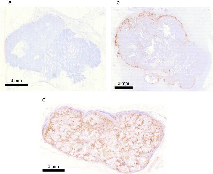
Programmed death-ligand 1 (PD-L1) plays important roles in the evasion of antitumor immunity. Because we observed the localization of PD-L1-positive (PD-L1) cells in the marginal region of triple-negative breast cancer (TNBC) specimens, we hypothesized that the marginal microenvironment of TNBC would involve the induction of PD-L1 cells.
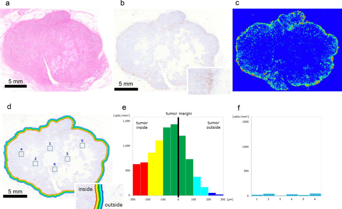
One hundred and one TNBC surgical specimens were examined. We performed immunohistochemical (IHC) studies of PD-L1, CD68, CD8, and pan-cytokeratin in these specimens. We analyzed the localization of IHC-positive cells and the distance between these cells by histological spatial analysis.
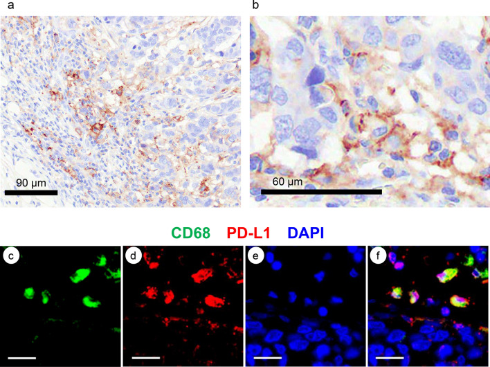
In 30.7% of TNBC specimens, PD-L1 cells were located in the marginal region. Approximately three PD-L1 cells accumulated around a single TNBC cell. Most PD-L1 cells were located within 50 μm of TNBC cells. PD-L1 cells were indicated to interact with TNBC cells in the marginal region. PD-L1CD68 cells were located in the marginal region, while CD68 macrophages (MΦs) were observed either in the marginal region or the core region. PD-L1 expression in MΦs was induced in the marginal region. The colocalization of CD8 T cells in the marginal region indicates that PD-L1 expression in MΦs would be induced by interaction with CD8 T cells. Because CD8 T cells are positive for CCL2, CCL2 may induce PD-L1 expression in MΦs.
At the marginal microenvironment of TNBC, PD-L1 expression would be induced in MΦs by interaction with CD8 T cells through CCL2. The interaction between PD-L1 MΦs and TNBC cells would facilitate the growth of TNBC under antitumor immunity. These interactions would be potential targets for restoring antitumor immunity and suppressing TNBC progression.
程序性死亡配体 1(PD-L1)在逃避抗肿瘤免疫中发挥重要作用。由于我们观察到三阴性乳腺癌(TNBC)标本中 PD-L1 阳性(PD-L1)细胞位于边缘区域,我们假设 TNBC 的边缘微环境将涉及 PD-L1 细胞的诱导。
检查了 101 例 TNBC 手术标本。我们对这些标本进行了 PD-L1、CD68、CD8 和细胞角蛋白的免疫组织化学(IHC)研究。我们通过组织学空间分析分析了 IHC 阳性细胞的定位和这些细胞之间的距离。
在 30.7%的 TNBC 标本中,PD-L1 细胞位于边缘区域。大约三个 PD-L1 细胞聚集在单个 TNBC 细胞周围。大多数 PD-L1 细胞位于 TNBC 细胞 50μm 范围内。PD-L1 细胞被指示与边缘区域的 TNBC 细胞相互作用。PD-L1CD68 细胞位于边缘区域,而 CD68 巨噬细胞(MΦ)则位于边缘区域或核心区域。MΦ 中的 PD-L1 表达在边缘区域被诱导。边缘区域 CD8 T 细胞的共定位表明,MΦ 中的 PD-L1 表达将通过与 CD8 T 细胞的相互作用而被诱导。由于 CD8 T 细胞对 CCL2 呈阳性,CCL2 可能诱导 MΦ 中的 PD-L1 表达。
在 TNBC 的边缘微环境中,通过 CCL2 与 CD8 T 细胞的相互作用,PD-L1 在 MΦ 中被诱导表达。PD-L1 MΦ 与 TNBC 细胞之间的相互作用将促进抗肿瘤免疫下 TNBC 的生长。这些相互作用可能成为恢复抗肿瘤免疫和抑制 TNBC 进展的潜在靶点。