Department of Pathology, Yamagata University Faculty of Medicine, Yamagata, Japan.
Department of Otolaryngology, Head and Neck Surgery, Yamagata University Faculty of Medicine, Yamagata, Japan.
J Clin Exp Hematop. 2024;64(3):223-231. doi: 10.3960/jslrt.24040.
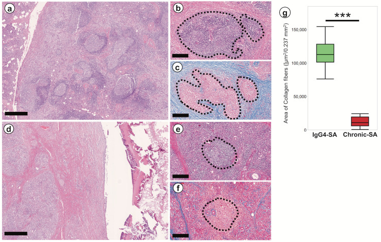
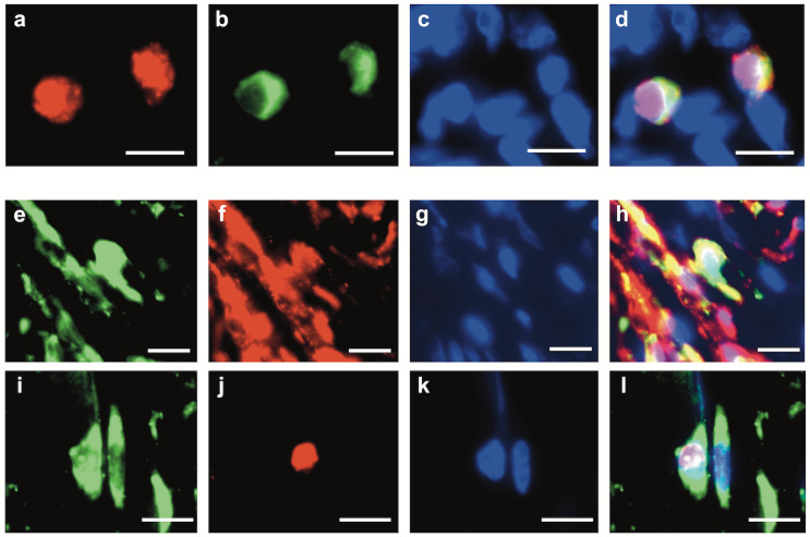
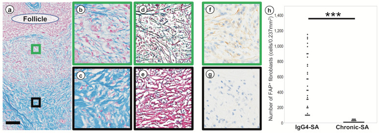
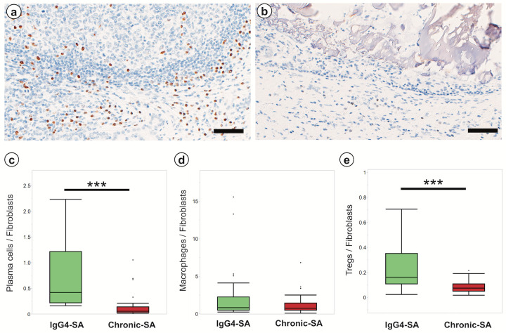
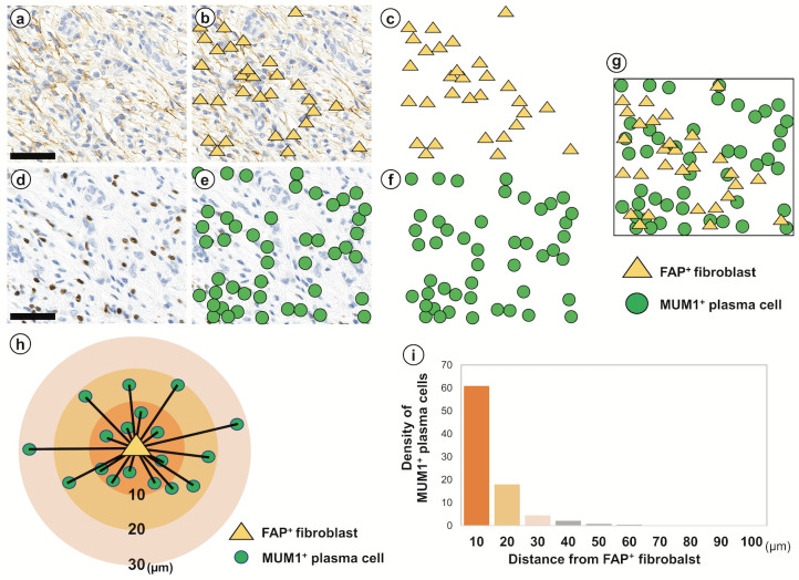
IgG4-related sialadenitis (IgG4-SA) is one of the IgG4-related disease. The histological features of IgG4-SA include dense lymphoplasmacytic infiltrates and fibrosis. This study aimed to reveal the involvement of plasma cells in the development of fibrosis and the mechanism underlying fibrosis in IgG4-SA. Hematoxylin-eosin staining, Azan staining, silver staining, and immunohistochemistry (IHC) were performed on IgG4-SA and chronic sialadenitis specimens, and theses samples were analyzed by image analysis software. Histological spatial analysis was used to analyze the localization of IHC-positive cells and the distances between these cells. In the IgG4-SA group, many secondary lymphoid follicles with germinal centers were found, and many collagen fibers developed around these germinal centers. Collagen fibers composed mainly of type I collagen was abundant at sites away from secondary lymphoid follicles, and reticular fibers composed of type III collagen was abundant near secondary lymphoid follicles. Many FAP fibroblasts and MUM1 plasma cells were localized near secondary lymphoid follicles. Histological spatial analysis demonstrated that 90.4% of MUM1 plasma cells accumulated within 20 µm of FAP fibroblasts. Multiple immunofluorescence assays revealed that MUM1 plasma cells expressed platelet-derived growth factor (PDGF) β, and FAP fibroblasts expressed PDGF receptor (PDGFR) β and pSTAT3 in IgG4-SA. We have shown that fibrosis is localized around secondary lymphoid follicles and that fibroblasts are activated by plasma cells via PDGF/PDGFR signaling in IgG4-SA.
IgG4 相关涎腺炎(IgG4-SA)是 IgG4 相关疾病之一。IgG4-SA 的组织学特征包括密集的淋巴浆细胞浸润和纤维化。本研究旨在揭示浆细胞在纤维化发展中的作用以及 IgG4-SA 纤维化的机制。对 IgG4-SA 和慢性涎腺炎标本进行苏木精-伊红染色、阿赞染色、银染色和免疫组织化学(IHC)染色,并通过图像分析软件对这些样本进行分析。组织学空间分析用于分析 IHC 阳性细胞的定位和这些细胞之间的距离。在 IgG4-SA 组中,发现了许多具有生发中心的次级淋巴滤泡,并且在这些生发中心周围形成了许多胶原纤维。远离次级淋巴滤泡的部位富含主要由 I 型胶原组成的胶原纤维,而靠近次级淋巴滤泡的部位富含由 III 型胶原组成的网状纤维。许多 FAP 成纤维细胞和 MUM1 浆细胞定位于次级淋巴滤泡附近。组织学空间分析表明,90.4%的 MUM1 浆细胞积聚在 FAP 成纤维细胞 20µm 范围内。多重免疫荧光检测显示,MUM1 浆细胞表达血小板衍生生长因子(PDGF)β,而 FAP 成纤维细胞在 IgG4-SA 中表达 PDGF 受体(PDGFR)β和 pSTAT3。我们已经表明,纤维化定位于次级淋巴滤泡周围,并且成纤维细胞通过 IgG4-SA 中的 PDGF/PDGFR 信号被浆细胞激活。