Department of Reproductive Medicine and Infertility, First IVF Clinic, Dubai Healthcare City, Dubai, UAE. (Dr. Madnani).
Department of Minimal Invasive Gynecological Surgery, Parikh Super Specialty Hospital, Ahmedabad, Gujarat, India. (Dr. Sonara).
CRSLS. 2023 Oct 6;10(3). doi: 10.4293/CRSLS.2023.00029. eCollection 2023 Jul-Sep.
Endometriosis originating in mesonephric cyst is unusual and with unknown prevalence. Endometriotic lesion in vestigial remnant of wolffian duct (mesonephric cyst) is exceptional. In the extended literature review only three cases have been reported in animal studies, and our case reported here is the first in human beings. We present a case of mesonephric cyst endometrioma in a 37-year-old patient who was referred for severe dysmenorrhea, long duration pelvic and back pain, subfertility, severe dyspareunia, and groin discomfort. The patient underwent laparoscopic removal and we performed a literature review to gain insight about the origin and surgical management of an atypical site endometriosis.
Case report presentation rests on information obtained from the patient database. We performed the literature review using a Medline search with the keywords: mesonephric cyst endometriosis, atypical location of endometriosis in vestigial remnant in wolffian duct, and Gartner duct cyst endometrioma.
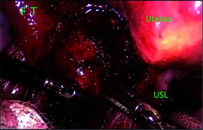
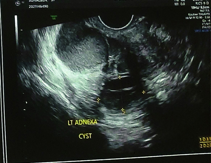
On physical examination, fullness and tenderness in left adnexa and lateral vaginal wall fullness on left side with restricted mobility of uterus was noted. Based on the examination and imaging the left ovarian cyst and mesonephric cyst were suspected. Surgical exploration revealed the left hemorrhagic cyst with deep infiltrating endometriosis involving left ureter and left uterosacral ligament with mesonephric cyst endometriosis. The review of literature revealed three cases where ectopic endometrial tissue in mesonephric cyst remnant was found in female dogs.
Mesonephric cyst endometrioma, although rare, can be a representative of extensive endometriosis. This case highlights an importance of careful clinical examination, correlation of patient symptoms with examination and imaging, and successful laparoscopic management of an atypical location endometriotic lesions. We completed the literature review on successful surgical management of such cases.
中肾管来源的子宫内膜异位症并不常见,其患病率也未知。残遗的沃尔夫管(中肾管)内的子宫内膜异位病变则更为罕见。在广泛的文献回顾中,仅在动物研究中报道了三例,而我们在此报告的病例是人类中的首例。我们报告了一例 37 岁患者的中肾管囊型子宫内膜异位症,该患者因严重痛经、长时间盆腔和背部疼痛、不孕、严重性交困难和腹股沟不适而就诊。患者接受了腹腔镜切除手术,我们进行了文献回顾,以深入了解非典型部位子宫内膜异位症的起源和手术治疗方法。
病例报告介绍基于从患者数据库中获得的信息。我们使用 Medline 搜索,关键词为:中肾管囊肿子宫内膜异位症、残遗沃尔夫管中子宫内膜异位症的非典型位置、以及 Gartner 管囊肿子宫内膜异位症,进行文献回顾。
体格检查时,左侧附件饱满和触痛,左侧侧阴道壁饱满,子宫活动受限。根据检查和影像学结果,怀疑左侧卵巢囊肿和中肾管囊肿。手术探查显示左侧出血性囊肿,伴有深部浸润性子宫内膜异位症,累及左侧输尿管和左侧子宫骶韧带,并伴有中肾管囊肿子宫内膜异位症。文献回顾显示,在雌性犬中发现了三个异位子宫内膜组织位于中肾管残遗的病例。
虽然中肾管囊型子宫内膜异位症罕见,但它可能是广泛子宫内膜异位症的代表。该病例强调了仔细的临床检查、患者症状与检查和影像学的相关性以及对非典型部位子宫内膜异位病变的成功腹腔镜管理的重要性。我们完成了对这些病例成功手术治疗的文献综述。