Yang Hao, Ping Xiyuan, Cui Yilei, Zheng Sifan, Shentu Xingchao
Department of Ophthalmology, The Second Affiliated Hospital of Zhejiang University School of Medicine; Zhejiang Provincial Key Lab of Ophthalmology, Hangzhou, China.
GKT School of Medical Education, King's College London, London, UK.
Adv Ophthalmol Pract Res. 2022 Oct 15;3(1):15-22. doi: 10.1016/j.aopr.2022.09.002. eCollection 2023 Feb-Mar.
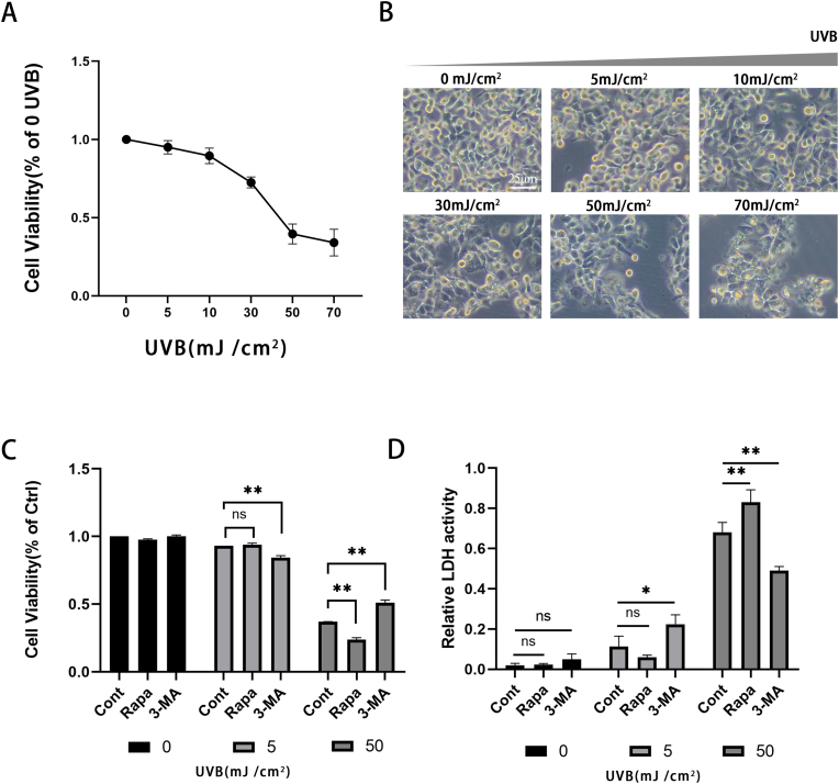
This study compared the role of autophagy regulators Rapamycin and 3-MA in oxidative damage and apoptosis of human lens epithelial cells (HLECs) caused by two doses of Ultraviolet Radiation B (UVB).
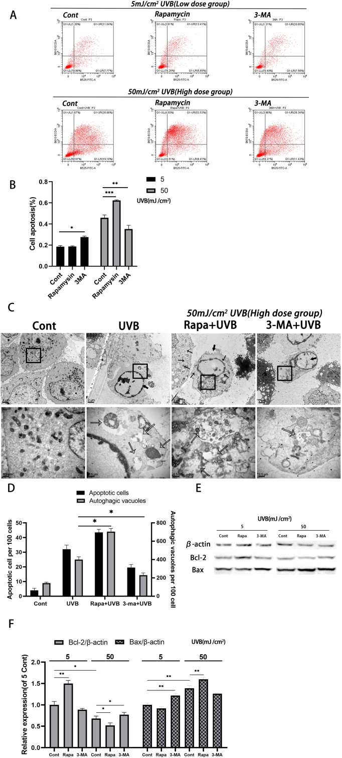
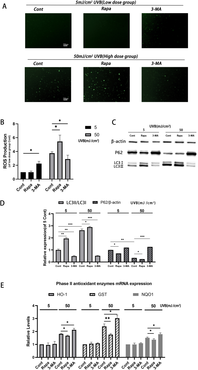
HLECs were irradiated with UVB, and two doses of UVB damage models were constructed. After treatment with autophagy regulators, cell damage tests such as CCK-8, LDH activity, and Ros detection were performed. Western blotting was used to detect the levels of autophagy-related proteins and apoptosis-related proteins. Quantitative real-time PCR (RT-qPCR) was used to detect the mRNA leve of secondary antioxidant enzymes.Flow cytometry was used to examine cell viability and apoptosis. Finally, the proportion of autophagy and apoptosis was observed by electron microscope.
Autophagy inhibitor 3-MA promoted oxidative damage and apoptosis of HLECs at low doses of UVB (5 mJ/cm2), which corresponds to 1.3 h of exposure to sunlight in human eyes. Under the high dose of UVB (50mJ/cm2), which is equivalent to 13 h of exposure to sunlight in human eyes, the autophagy inducer Rapamycin caused more extensive oxidative damage and apoptosis of HLECs. 3-MA was able to reduce this damage, indicating that moderate autophagy is necessary for HLECs to cope with mild oxidative stress. For high dose UVB-induced oxidative stress, the use of 3-MA inhibiting autophagy is more beneficial to reduce cell damage and apoptosis. The mechanisms include degradation of damaged organelles, regulation of the expression of antioxidant enzymes HO-1, NQO1, GCS and regulation of apoptosis-related proteins.
Autophagy played different roles in HLECs oxidative stress induced by two doses of UVB. It provides new ideas for reducing oxidative damage and apoptosis of HLECs to prevent or delay the progression of age-related cataract (ARC).
本研究比较了自噬调节剂雷帕霉素和3-甲基腺嘌呤(3-MA)在两剂量紫外线B(UVB)引起的人晶状体上皮细胞(HLECs)氧化损伤和凋亡中的作用。
用UVB照射HLECs,构建两剂量UVB损伤模型。用自噬调节剂处理后,进行CCK-8、乳酸脱氢酶(LDH)活性和活性氧(Ros)检测等细胞损伤试验。采用蛋白质免疫印迹法检测自噬相关蛋白和凋亡相关蛋白的水平。采用定量实时聚合酶链反应(RT-qPCR)检测二级抗氧化酶的mRNA水平。采用流式细胞术检测细胞活力和凋亡情况。最后,通过电子显微镜观察自噬和凋亡的比例。
自噬抑制剂3-MA在低剂量UVB(5 mJ/cm2,相当于人眼暴露于阳光下1.3小时)时促进HLECs的氧化损伤和凋亡。在高剂量UVB(50 mJ/cm2,相当于人眼暴露于阳光下13小时)下,自噬诱导剂雷帕霉素导致HLECs更广泛的氧化损伤和凋亡。3-MA能够减轻这种损伤,表明适度自噬对于HLECs应对轻度氧化应激是必要的。对于高剂量UVB诱导的氧化应激,使用3-MA抑制自噬更有利于减少细胞损伤和凋亡。其机制包括受损细胞器的降解、抗氧化酶HO-1、NQO1、谷胱甘肽半胱氨酸合成酶(GCS)表达的调节以及凋亡相关蛋白的调节。
自噬在两剂量UVB诱导的HLECs氧化应激中发挥不同作用。这为减少HLECs的氧化损伤和凋亡以预防或延缓年龄相关性白内障(ARC)的进展提供了新思路。