Haem Rahimi Muzhda, Bidar Frank, Lukaszewicz Anne-Claire, Garnier Lorna, Payen-Gay Léa, Venet Fabienne, Monneret Guillaume
Hospices Civils de Lyon, Guillaume Monneret - Immunology Laboratory, Hôpital E. Herriot, Lyon, France.
Université de Lyon, EA 7426 "Pathophysiology of Injury-Induced Immunosuppression", Université Claude Bernard Lyon_1, Lyon, France.
Ann Intensive Care. 2023 Oct 17;13(1):102. doi: 10.1186/s13613-023-01204-y.
Understanding the mechanisms underlying immune dysregulation in sepsis is a major challenge in developing more individualized therapy, as early and persistent inflammation, as well as immunosuppression, play a significant role in pathophysiology. As part of the antimicrobial response, neutrophils can release extracellular traps (NETs) which neutralize and kill microorganisms. However, excessive NETs formation may also contribute to pathogenesis, tissue damage and organ dysfunction. Recently, a novel automated assay has been proposed for the routine measurement of nucleosomes H3.1 (fundamental units of chromatin) that are released during NETs formation. The aim of the present study was to measure nucleosome levels in 151 septic shock patients (according to sepsis-3 definition) and to determine association with mortality.
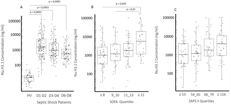
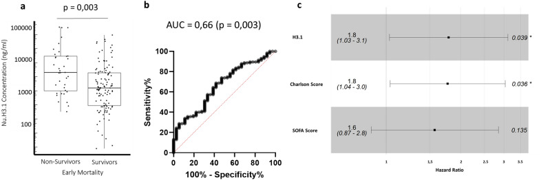
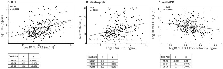
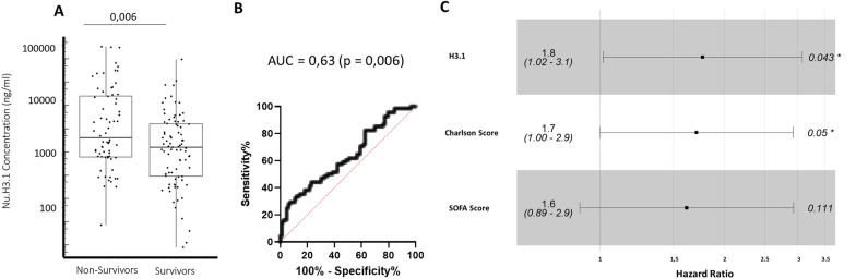
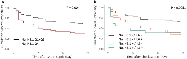
The nucleosome H3.1 levels (as determined by a chemiluminescence immunoassay performed on an automated immunoanalyzer system) were markedly and significantly elevated at all-time points in septic shock patients compared to the control group. Immunological parameters indicated tremendous early inflammation (IL-6 = 1335 pg/mL at day 1-2) along with marked immunosuppression (e.g., mHLA-DR = 3853 AB/C and CD4 = 338 cell /µL at day 3-4). We found significantly positive correlation between nucleosome levels and organ failure and severity scores, IL-6 concentrations and neutrophil count. Significantly higher values (day 1-2 and 3-4) were measured in non-survivor patients (28-day mortality). This association was still significant after multivariate analysis and was more pronounced with highest concentration. Early (day 1-2) increased nucleosome levels were also independently associated with 5-day mortality. At day 6-8, persistent elevated nucleosome levels were negatively correlated to mHLA-DR values.
This study reports a significant elevation of nucleosome in patients during a one-week follow-up. The nucleosome levels showed correlation with neutrophil count, IL-6 and were found to be independently associated with mortality assessed at day 5 or 28. Therefore, nucleosome concentration seems to be a promising biomarker for detecting hyper-inflammatory phenotype upon a patient's admission. Additional investigations are required to evaluate the potential association between sustained elevation of nucleosome and sepsis-induced immunosuppression.
了解脓毒症中免疫失调的潜在机制是开发更具个性化治疗方法的一项重大挑战,因为早期和持续的炎症以及免疫抑制在病理生理学中起着重要作用。作为抗菌反应的一部分,中性粒细胞可释放细胞外陷阱(NETs),其可中和并杀死微生物。然而,过量的NETs形成也可能导致发病机制、组织损伤和器官功能障碍。最近,一种新型自动化检测方法被提出用于常规测量NETs形成过程中释放的核小体H3.1(染色质的基本单位)。本研究的目的是测量151例感染性休克患者(根据脓毒症-3定义)的核小体水平,并确定其与死亡率的关联。
与对照组相比,感染性休克患者在所有时间点的核小体H3.1水平(通过在自动化免疫分析仪系统上进行的化学发光免疫分析测定)均显著升高。免疫参数表明存在严重的早期炎症(第1 - 2天IL-6 = 1335 pg/mL)以及明显的免疫抑制(例如,第3 - 4天mHLA-DR = 3853 AB/C且CD4 = 338细胞/µL)。我们发现核小体水平与器官衰竭、严重程度评分、IL-6浓度和中性粒细胞计数之间存在显著正相关。在非存活患者(28天死亡率)中测量到的值(第1 - 2天和第3 - 4天)显著更高。多因素分析后这种关联仍然显著,且在浓度最高时更为明显。早期(第1 - 2天)核小体水平升高也与5天死亡率独立相关。在第6 - 8天,持续升高的核小体水平与mHLA-DR值呈负相关。
本研究报告了患者在为期一周的随访期间核小体显著升高。核小体水平与中性粒细胞计数、IL-6相关,并且被发现与第5天或第28天评估的死亡率独立相关。因此,核小体浓度似乎是患者入院时检测高炎症表型的一个有前景的生物标志物。需要进一步研究来评估核小体持续升高与脓毒症诱导的免疫抑制之间的潜在关联。