• 文献检索
  • 文档翻译
  • 深度研究
  • 学术资讯
  • Suppr Zotero 插件Zotero 插件
  • 邀请有礼
  • 套餐&价格
  • 历史记录
应用&插件
Suppr Zotero 插件Zotero 插件浏览器插件Mac 客户端Windows 客户端微信小程序
定价
高级版会员购买积分包购买API积分包
服务
文献检索文档翻译深度研究API 文档MCP 服务
关于我们
关于 Suppr公司介绍联系我们用户协议隐私条款
关注我们

Suppr 超能文献

核心技术专利:CN118964589B侵权必究
粤ICP备2023148730 号-1Suppr @ 2026

文献检索

告别复杂PubMed语法,用中文像聊天一样搜索,搜遍4000万医学文献。AI智能推荐,让科研检索更轻松。

立即免费搜索

文件翻译

保留排版,准确专业,支持PDF/Word/PPT等文件格式,支持 12+语言互译。

免费翻译文档

深度研究

AI帮你快速写综述,25分钟生成高质量综述,智能提取关键信息,辅助科研写作。

立即免费体验

后交通动脉瘤的产前特征:一例报告。

Prenatal features of aneurysm of the posterior cerebral artery: a case report.

机构信息

Department of Obstetrics and Gynecology, Sawanpracharak Hospital, Nakhon Sawan, Thailand.

Department of Obstetrics and Gynecology, Faculty of Medicine, Chiang Mai University, Chiang Mai 50200, Chiang Mai, Thailand.

出版信息

BMC Pregnancy Childbirth. 2023 Oct 17;23(1):734. doi: 10.1186/s12884-023-06056-9.

DOI:10.1186/s12884-023-06056-9
PMID:37848838
原文链接:https://pmc.ncbi.nlm.nih.gov/articles/PMC10583320/
Abstract

BACKGROUND

Fetal cerebral aneurysm other than aneurysm of vein of Galen aneurysmal malformation (VGAM) is extremely rare. This report describes prenatal features of aneurysm of the posterior cerebral artery (APCA) with rapid progression and its natural intrauterine course of the disease, which has never been reported.

CASE PRESENTATION

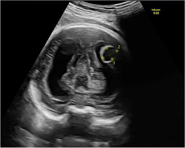
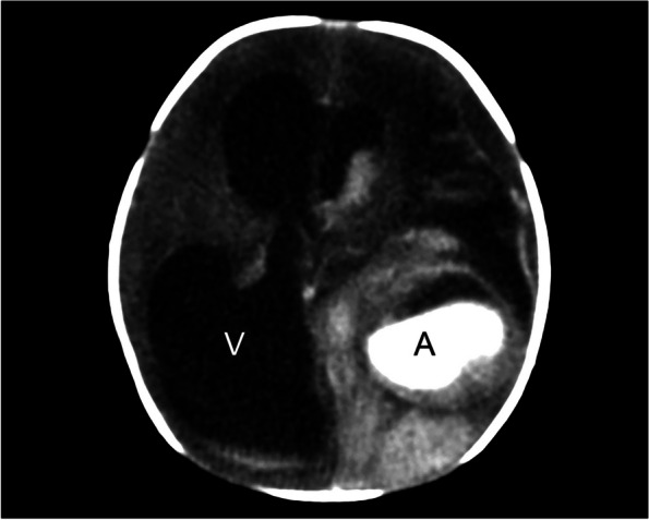
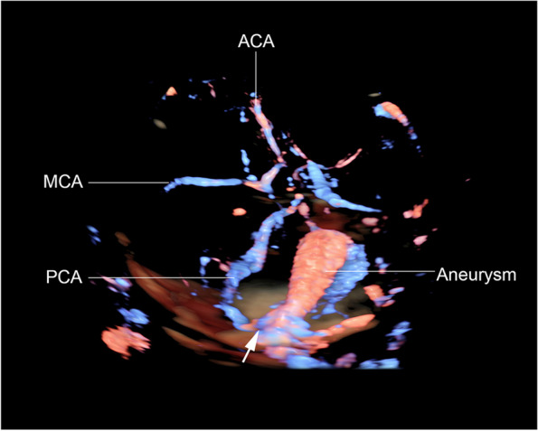
This is the first report of prenatal features of APCA, detected at 34-36 weeks of gestation, simulating choroid plexus cyst or arachnoid cyst. The diagnosis was based on color flow ultrasound with tracing along the course of cerebral arteries. Also, rendered 3D color flow ultrasound was helpful in demonstrating course of the vessels feeding the aneurysm and supporting the diagnosis. The aneurysm showed nature of rapidly progressive changes, leading to leakage resulting in intracerebral and intraventricular hemorrhage as well as high output state associated with anemia. Prenatal diagnosis and management are very challenging. This case ended up with planned delivery at 37 weeks, giving birth to a surviving male newborn, weighing 2600 g. The neonatal CT brain scans and CTA confirmed the prenatal findings. The prognosis was relatively poor because of extensive intracerebral hemorrhage with severe hydrocephalus and brain midline shift. The couple opted for neonatal palliative care without neurosurgical correction.

CONCLUSION

This study demonstrate that the most important tool for prenatal diagnosis is color Doppler ultrasound, which will demonstrate turbulent blood flow. Three-dimension color Doppler ultrasound is helpful in supporting the diagnosis. The case presented here suggests that the disease has a natural course of rapid progression and massive brain destruction or high output congestive heart failure can be expected.

摘要

背景

除了静脉瘤样畸形(VGAM)的脑动脉瘤外,胎儿脑动脉瘤极为罕见。本报告描述了一种快速进展的大脑后动脉动脉瘤(APCA)的产前特征及其疾病的自然宫内过程,这是从未报道过的。

病例介绍

这是首例产前大脑后动脉动脉瘤的特征报告,在 34-36 孕周时被检测到,模拟脉络丛囊肿或蛛网膜囊肿。该诊断基于彩色血流超声,沿着大脑动脉的走行进行追踪。此外,渲染的 3D 彩色血流超声有助于显示为动脉瘤供血的血管的走行,支持诊断。动脉瘤显示出快速进展变化的特征,导致漏出,引起脑内和脑室内出血以及与贫血相关的高输出状态。产前诊断和管理极具挑战性。本例最终在 37 孕周时计划分娩,生下一名存活的男婴,体重 2600 克。新生儿 CT 脑扫描和 CTA 证实了产前发现。由于广泛的脑内出血、严重的脑积水和大脑中线移位,预后相对较差。夫妇选择新生儿姑息治疗,而不进行神经外科矫正。

结论

本研究表明,产前诊断最重要的工具是彩色多普勒超声,它将显示湍流的血流。三维彩色多普勒超声有助于支持诊断。本病例提示该疾病具有快速进展和大量脑破坏或高输出充血性心力衰竭的自然病程。

https://cdn.ncbi.nlm.nih.gov/pmc/blobs/dd6c/10583320/746cce1ee0dc/12884_2023_6056_Fig4_HTML.jpg
https://cdn.ncbi.nlm.nih.gov/pmc/blobs/dd6c/10583320/403dbaba6980/12884_2023_6056_Fig1_HTML.jpg
https://cdn.ncbi.nlm.nih.gov/pmc/blobs/dd6c/10583320/bcfc9ac749ac/12884_2023_6056_Fig2_HTML.jpg
https://cdn.ncbi.nlm.nih.gov/pmc/blobs/dd6c/10583320/87e41249a770/12884_2023_6056_Fig3_HTML.jpg
https://cdn.ncbi.nlm.nih.gov/pmc/blobs/dd6c/10583320/746cce1ee0dc/12884_2023_6056_Fig4_HTML.jpg

相似文献

1
Prenatal features of aneurysm of the posterior cerebral artery: a case report.后交通动脉瘤的产前特征:一例报告。
BMC Pregnancy Childbirth. 2023 Oct 17;23(1):734. doi: 10.1186/s12884-023-06056-9.
2
Prenatal diagnosis of a vein of Galen aneurysmal malformation using color Doppler ultrasound: a case report.使用彩色多普勒超声对大脑大静脉动脉瘤样畸形进行产前诊断:一例报告
Bol Asoc Med P R. 2013;105(1):32-5.
3
Prenatal diagnosis of an aneurysm of the vein of galen by three-dimensional power and color Doppler ultrasonography.三维能量及彩色多普勒超声对大脑大静脉瘤的产前诊断
Clin Med Insights Case Rep. 2012;5:77-80. doi: 10.4137/CCRep.S9597. Epub 2012 May 24.
4
Antenatal diagnosis and prognostic factors of aneurysmal malformation of the vein of Galen: A case report and literature review.大脑大静脉动脉瘤样畸形的产前诊断及预后因素:1例病例报告并文献复习
Medicine (Baltimore). 2017 Jul;96(30):e7483. doi: 10.1097/MD.0000000000007483.
5
Vein of Galen aneurysm: antenatal diagnosis: a case report.大脑大静脉动脉瘤:产前诊断:病例报告
J Pak Med Assoc. 2005 Oct;55(10):455-6.
6
Prenatal diagnosis of vein of Galen aneurysmal malformation: report of two cases with proposal for prognostic indices.大脑大静脉瘤样畸形的产前诊断:两例报告及预后指标建议
Prenat Diagn. 1997 Oct;17(10):972-7.
7
Aneurysm of the vein of Galen: prenatal diagnosis and perinatal management.大脑大静脉瘤:产前诊断与围产期管理
Am J Perinatol. 1996 Nov;13(8):503-6. doi: 10.1055/s-2007-994437.
8
The significance of 3D power Doppler in prenatal diagnosis and the evaluation of the anatomical structure of vein of Galen aneurysmatic malformation: case report.三维能量多普勒在产前诊断及大脑大静脉动脉瘤样畸形静脉解剖结构评估中的意义:病例报告
Clin Exp Obstet Gynecol. 2013;40(2):300-3.
9
Diagnosis of Vein of Galen aneurysmal malformation using fetal MRI.使用胎儿 MRI 诊断大脑大静脉瘤样畸形。
J Magn Reson Imaging. 2017 Nov;46(5):1535-1539. doi: 10.1002/jmri.25478. Epub 2016 Sep 30.
10
Ultrasound Prenatal Diagnosis and Emergency Interventional Radiologic Therapy of Galen Aneurysmal Malformation in a Newborn.新生儿大脑大静脉动脉瘤样畸形的超声产前诊断与急诊介入放射治疗
Maedica (Bucur). 2016 Dec;11(4):334-340.

本文引用的文献

1
Duplicated fetal posterior cerebral artery in a patient with a ruptured fetal posterior cerebral artery aneurysm: a cerebrovascular variant.胎儿大脑后动脉重复畸形合并胎儿大脑后动脉瘤破裂1例:一种脑血管变异
Surg Radiol Anat. 2023 Jan;45(1):35-38. doi: 10.1007/s00276-022-03052-0. Epub 2022 Dec 9.
2
Mechanical thrombectomy for acute fetal posterior cerebral artery occlusion with a hidden unruptured cerebral aneurysm: illustrative case.机械取栓治疗急性胎儿大脑后动脉闭塞合并隐匿性未破裂脑动脉瘤:病例说明
J Neurosurg Case Lessons. 2022 Aug 29;4(9):CASE22291. doi: 10.3171/CASE22291.
3
Treatment of a ruptured fetal-type posterior communicating aneurysm with a combined approach using the new contour neurovascular system.
应用新型轮廓神经血管系统联合治疗破裂胎儿型后交通动脉瘤。
BMJ Case Rep. 2022 Apr 19;15(4):e248065. doi: 10.1136/bcr-2021-248065.
4
Vein of Galen Aneurysmal Malformation.大脑大静脉动脉瘤样畸形
Am J Obstet Gynecol. 2020 Dec;223(6):B27-B29. doi: 10.1016/j.ajog.2020.08.181. Epub 2020 Nov 7.
5
Uncommon second-trimester presentation of vein of Galen malformation.妊娠中期罕见的大脑大静脉畸形表现。
Ultrasound Obstet Gynecol. 2018 Mar;51(3):421-423. doi: 10.1002/uog.17462.
6
Vein of Galen aneurysmal malformation (VGAM) in the fetus: retrospective analysis of perinatal prognostic indicators in a two-center series of 49 cases.胎儿静脉窦瘤样畸形(VGAM):两中心 49 例系列回顾性分析围产预后指标。
Ultrasound Obstet Gynecol. 2017 Aug;50(2):192-199. doi: 10.1002/uog.17224.
7
Risk profile of intracranial aneurysms: rupture rate is not constant after formation.颅内动脉瘤的风险特征:形成后破裂率并非恒定不变。
Stroke. 2011 Dec;42(12):3376-81. doi: 10.1161/STROKEAHA.111.625871. Epub 2011 Oct 6.
8
Pathogenesis, natural history, and treatment of unruptured intracranial aneurysms.未破裂颅内动脉瘤的发病机制、自然史及治疗
Mayo Clin Proc. 2004 Dec;79(12):1572-83. doi: 10.4065/79.12.1572.
9
Lethal progression of a fetal intracranial arteriovenous malformation.胎儿颅内动静脉畸形的致死性进展
J Ultrasound Med. 2003 Jun;22(6):645-8. doi: 10.7863/jum.2003.22.6.645.
10
Prenatal diagnosis of a vein of galen aneurysm.大脑大静脉动脉瘤的产前诊断。
Fetal Diagn Ther. 2003 Jan-Feb;18(1):36-40. doi: 10.1159/000066382.