• 文献检索
  • 文档翻译
  • 深度研究
  • 学术资讯
  • Suppr Zotero 插件Zotero 插件
  • 邀请有礼
  • 套餐&价格
  • 历史记录
应用&插件
Suppr Zotero 插件Zotero 插件浏览器插件Mac 客户端Windows 客户端微信小程序
定价
高级版会员购买积分包购买API积分包
服务
文献检索文档翻译深度研究API 文档MCP 服务
关于我们
关于 Suppr公司介绍联系我们用户协议隐私条款
关注我们

Suppr 超能文献

核心技术专利:CN118964589B侵权必究
粤ICP备2023148730 号-1Suppr @ 2026

文献检索

告别复杂PubMed语法,用中文像聊天一样搜索,搜遍4000万医学文献。AI智能推荐,让科研检索更轻松。

立即免费搜索

文件翻译

保留排版,准确专业,支持PDF/Word/PPT等文件格式,支持 12+语言互译。

免费翻译文档

深度研究

AI帮你快速写综述,25分钟生成高质量综述,智能提取关键信息,辅助科研写作。

立即免费体验

骨质疏松症的功能光声与超声评估:一项临床可行性研究。

Functional Photoacoustic and Ultrasonic Assessment of Osteoporosis: A Clinical Feasibility Study.

作者信息

Feng Ting, Zhu Yunhao, Morris Richard, Kozloff Kenneth M, Wang Xueding

机构信息

Department of Biomedical Engineering, University of Michigan Medical School, MI 48109, USA.

IF, LLC, WI 53589USA.

出版信息

BME Front. 2020 Oct 30;2020:1081540. doi: 10.34133/2020/1081540. eCollection 2020.

DOI:10.34133/2020/1081540
PMID:37849970
原文链接:https://pmc.ncbi.nlm.nih.gov/articles/PMC10521673/
Abstract

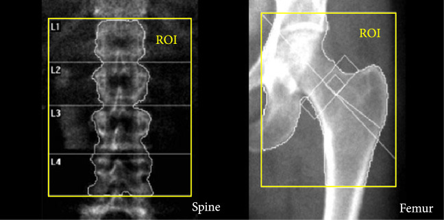
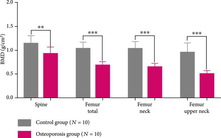
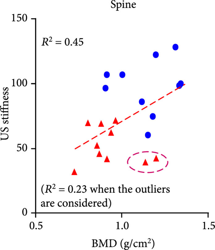
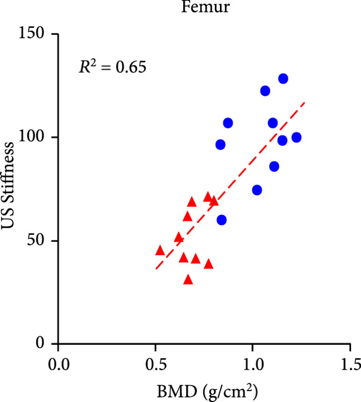
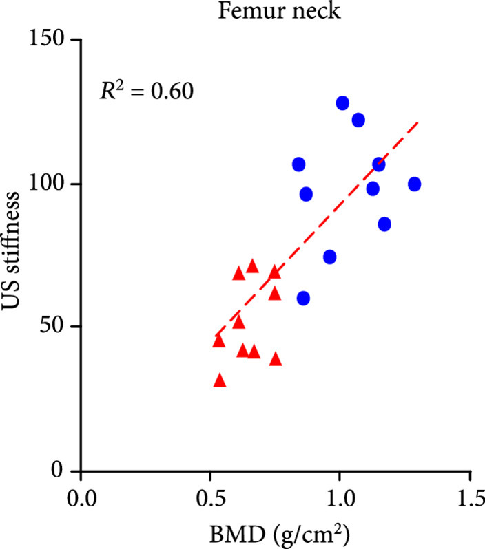
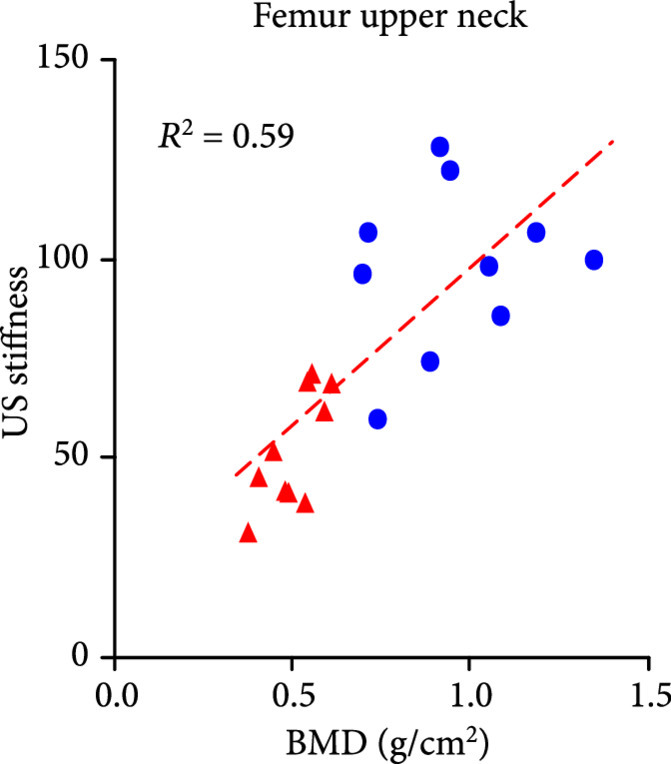
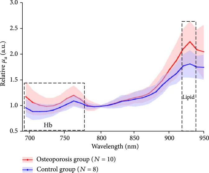
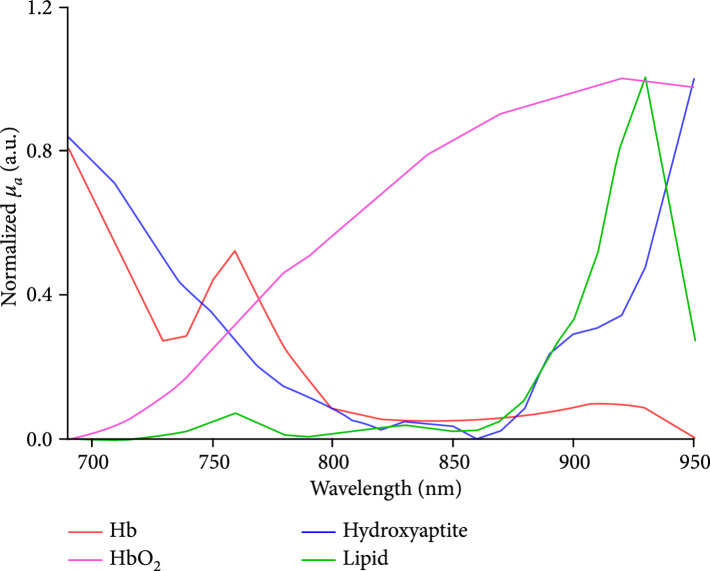
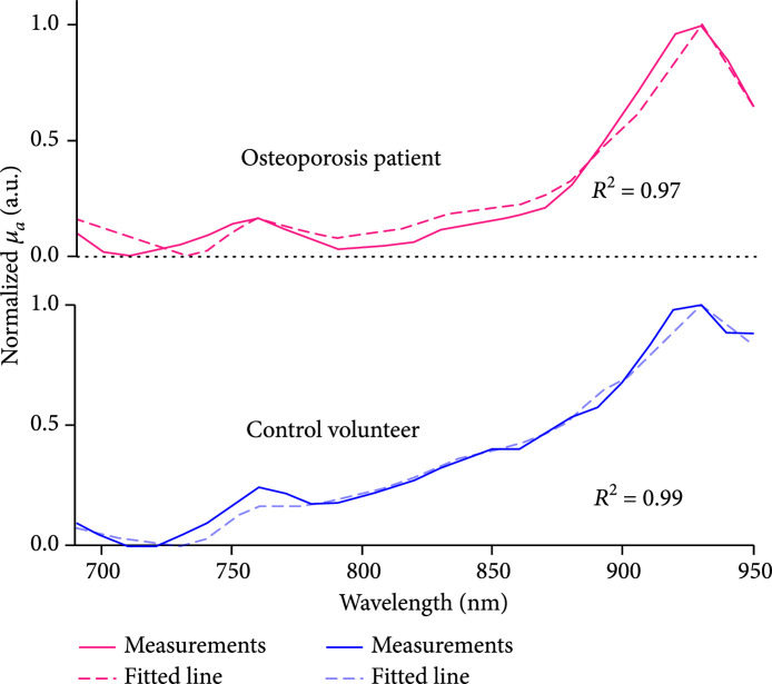
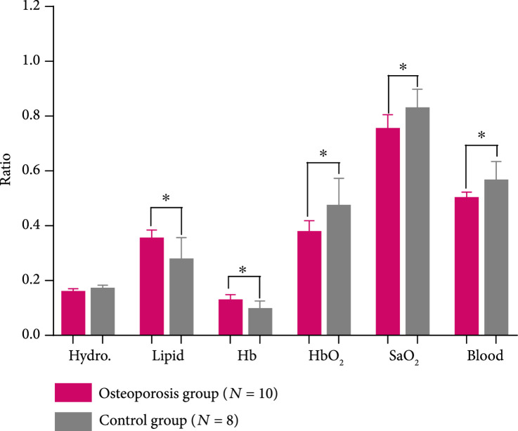
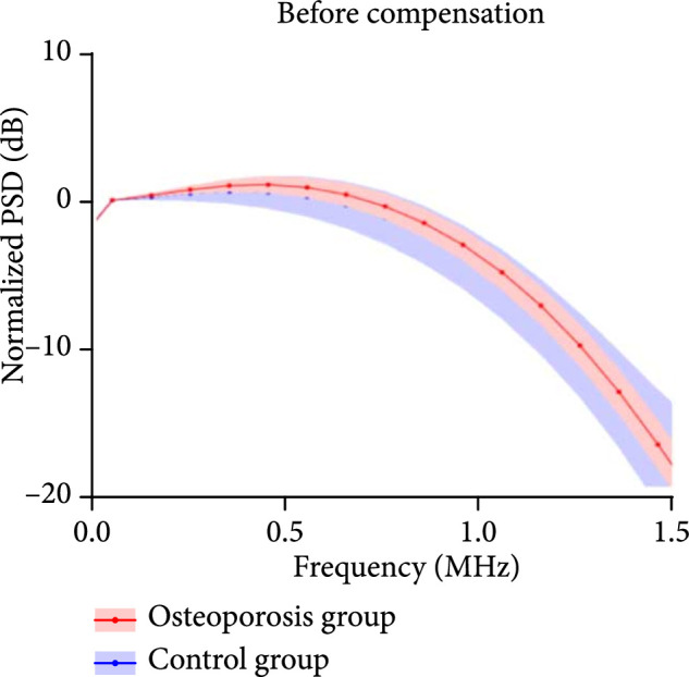
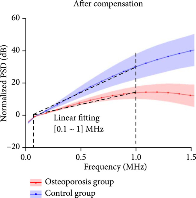
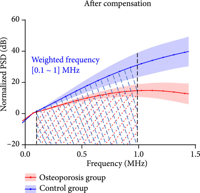
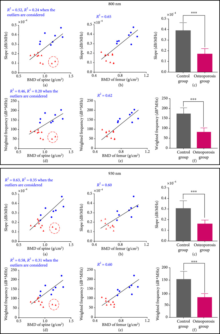
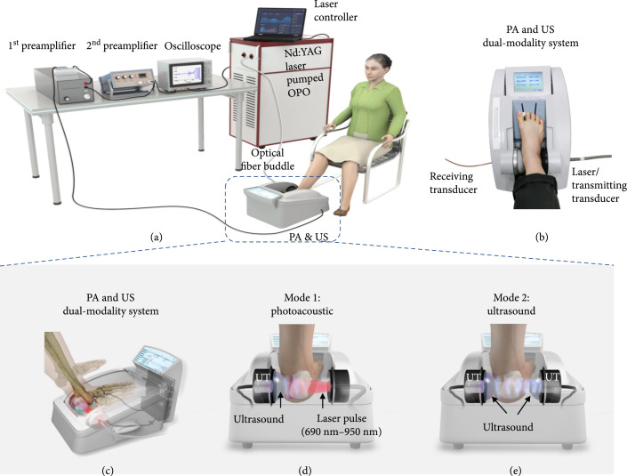
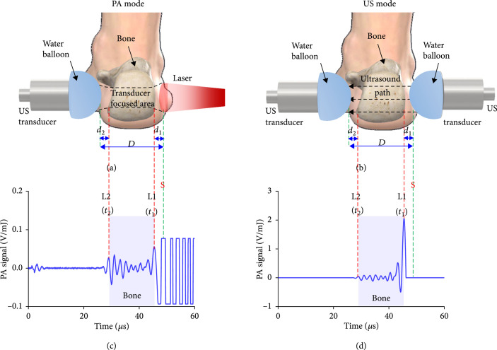
. To study the feasibility of combined functional photoacoustic (PA) and quantitative ultrasound (US) for diagnosis of osteoporosis based on the detection of chemical and microarchitecture (BMA) information in calcaneus bone. . Clinically available X-ray or US technologies for the diagnosis of osteoporosis do not report important parameters such as chemical information and BMA. With unique advantages, including good sensitivity to molecular and metabolic properties, PA bone assessment techniques hold a great potential for clinical translation. . By performing multiwavelength PA measurements, the chemical information in the human calcaneus bone, including mineral, lipid, oxygenated-hemoglobin, and deoxygenated-hemoglobin, were assessed. In parallel, by performing PA spectrum analysis, the BMA as an important bone physical property was quantified. An unpaired -test and a two-way ANOVA test were conducted to compare the outcomes from the two subject groups. . Multiwavelength PA measurement is capable of assessing the relative contents of several chemical components in the trabecular bone , including both minerals and organic materials such as oxygenated-hemoglobin, deoxygenated-hemoglobin, and lipid, which are relevant to metabolic activities and bone health. In addition, PA measurements of BMA show good correlations ( up to 0.65) with DEXA. Both the chemical and microarchitectural measurements from PA techniques can differentiate the two subject groups. . The results from this initial clinical study suggest that PA techniques, by providing additional chemical and microarchitecture information relevant to bone health, may lead to accurate and early diagnosis, as well as sensitive monitoring of the treatment of osteoporosis.

摘要

为了基于对跟骨化学和微观结构(骨矿物质密度)信息的检测,研究联合功能光声(PA)和定量超声(US)诊断骨质疏松症的可行性。临床可用的用于诊断骨质疏松症的X射线或超声技术并未报告诸如化学信息和骨矿物质密度等重要参数。光声骨评估技术具有独特优势,包括对分子和代谢特性具有良好的敏感性,在临床转化方面具有巨大潜力。通过进行多波长光声测量,评估了人跟骨中的化学信息,包括矿物质、脂质、氧合血红蛋白和脱氧血红蛋白。同时,通过进行光声光谱分析,对作为重要骨物理特性的骨矿物质密度进行了量化。进行了非配对t检验和双向方差分析以比较两个受试者组的结果。多波长光声测量能够评估小梁骨中几种化学成分的相对含量,包括矿物质和有机物质,如氧合血红蛋白、脱氧血红蛋白和脂质,这些与代谢活动和骨骼健康相关。此外,骨矿物质密度的光声测量与双能X线吸收法显示出良好的相关性(高达0.65)。光声技术的化学和微观结构测量都可以区分两个受试者组。这项初步临床研究的结果表明,光声技术通过提供与骨骼健康相关的额外化学和微观结构信息,可能导致对骨质疏松症的准确早期诊断以及对治疗的敏感监测。

https://cdn.ncbi.nlm.nih.gov/pmc/blobs/f933/10521673/82c54810d7ac/1081540.fig.007.jpg
https://cdn.ncbi.nlm.nih.gov/pmc/blobs/f933/10521673/19b57c00dd12/1081540.fig.001a.jpg
https://cdn.ncbi.nlm.nih.gov/pmc/blobs/f933/10521673/f53acb0366fa/1081540.fig.001b.jpg
https://cdn.ncbi.nlm.nih.gov/pmc/blobs/f933/10521673/f2c9380c900e/1081540.fig.001c.jpg
https://cdn.ncbi.nlm.nih.gov/pmc/blobs/f933/10521673/dbf4c11f7345/1081540.fig.001d.jpg
https://cdn.ncbi.nlm.nih.gov/pmc/blobs/f933/10521673/ecd4a6b5fdb4/1081540.fig.001e.jpg
https://cdn.ncbi.nlm.nih.gov/pmc/blobs/f933/10521673/2b41c2811391/1081540.fig.001f.jpg
https://cdn.ncbi.nlm.nih.gov/pmc/blobs/f933/10521673/bf84d5a7a82d/1081540.fig.002a.jpg
https://cdn.ncbi.nlm.nih.gov/pmc/blobs/f933/10521673/d795d01bac9a/1081540.fig.002b.jpg
https://cdn.ncbi.nlm.nih.gov/pmc/blobs/f933/10521673/a1f6c97bd292/1081540.fig.002c.jpg
https://cdn.ncbi.nlm.nih.gov/pmc/blobs/f933/10521673/2dd9a17aca6f/1081540.fig.002d.jpg
https://cdn.ncbi.nlm.nih.gov/pmc/blobs/f933/10521673/1ebe6a7ccb62/1081540.fig.003a.jpg
https://cdn.ncbi.nlm.nih.gov/pmc/blobs/f933/10521673/f22ca3e8ea83/1081540.fig.003b.jpg
https://cdn.ncbi.nlm.nih.gov/pmc/blobs/f933/10521673/6b014b48574d/1081540.fig.003c.jpg
https://cdn.ncbi.nlm.nih.gov/pmc/blobs/f933/10521673/b15284aca0ce/1081540.fig.004.jpg
https://cdn.ncbi.nlm.nih.gov/pmc/blobs/f933/10521673/b526ae1edf5a/1081540.fig.005.jpg
https://cdn.ncbi.nlm.nih.gov/pmc/blobs/f933/10521673/ed381dc091a5/1081540.fig.006.jpg
https://cdn.ncbi.nlm.nih.gov/pmc/blobs/f933/10521673/82c54810d7ac/1081540.fig.007.jpg

相似文献

1
Functional Photoacoustic and Ultrasonic Assessment of Osteoporosis: A Clinical Feasibility Study.骨质疏松症的功能光声与超声评估:一项临床可行性研究。
BME Front. 2020 Oct 30;2020:1081540. doi: 10.34133/2020/1081540. eCollection 2020.
2
The feasibility study of the transmission mode photoacoustic measurement of human calcaneus bone .人体跟骨骨传输模式光声测量的可行性研究
Photoacoustics. 2021 May 26;23:100273. doi: 10.1016/j.pacs.2021.100273. eCollection 2021 Sep.
3
Assessment of bone microarchitecture in chronic kidney disease: a comparison of 2D bone texture analysis and high-resolution peripheral quantitative computed tomography at the radius and tibia.评估慢性肾脏病患者的骨微结构:2 维骨纹理分析与桡骨和胫骨高分辨率外周定量 CT 的比较。
Calcif Tissue Int. 2010 Nov;87(5):385-91. doi: 10.1007/s00223-010-9402-z. Epub 2010 Aug 15.
4
The application of backscattered ultrasound and photoacoustic signals for assessment of bone collagen and mineral contents.背向散射超声和光声信号在评估骨胶原和矿物质含量中的应用。
Quant Imaging Med Surg. 2015 Feb;5(1):46-56. doi: 10.3978/j.issn.2223-4292.2014.11.11.
5
Characterization of bone microstructure using photoacoustic spectrum analysis.利用光声光谱分析对骨微观结构进行表征。
Opt Express. 2015 Sep 21;23(19):25217-24. doi: 10.1364/OE.23.025217.
6
Wavelet transform-based photoacoustic time-frequency spectral analysis for bone assessment.基于小波变换的骨评估光声时频光谱分析
Photoacoustics. 2021 Mar 10;22:100259. doi: 10.1016/j.pacs.2021.100259. eCollection 2021 Jun.
7
Characterization of multi-biomarkers for bone health assessment based on photoacoustic physicochemical analysis method.基于光声物理化学分析方法的骨健康评估多生物标志物表征
Photoacoustics. 2021 Dec 14;25:100320. doi: 10.1016/j.pacs.2021.100320. eCollection 2022 Mar.
8
People living with HIV have low trabecular bone mineral density, high bone marrow adiposity, and poor trabecular bone microarchitecture at the proximal femur.HIV 感染者的股骨近端存在骨小梁骨密度低、骨髓脂肪含量高和骨小梁微结构差的情况。
Osteoporos Int. 2022 Aug;33(8):1739-1753. doi: 10.1007/s00198-022-06405-y. Epub 2022 Apr 27.
9
Coregistered photoacoustic and ultrasonic signatures of early bone density variations.早期骨密度变化的共配准光声和超声特征。
J Biomed Opt. 2014 Mar;19(3):36015. doi: 10.1117/1.JBO.19.3.036015.
10
Computed tomography image analysis of the calcaneus in male osteoporosis.男性骨质疏松症跟骨的计算机断层扫描图像分析
Osteoporos Int. 2002 Jan;13(1):33-41. doi: 10.1007/s198-002-8335-4.

引用本文的文献

1
Theoretical and experimental study of attenuation in cancellous bone.松质骨衰减的理论和实验研究。
J Biomed Opt. 2024 Jan;29(Suppl 1):S11526. doi: 10.1117/1.JBO.29.S1.S11526. Epub 2024 Mar 19.
2
Development of a semi-anthropomorphic photoacoustic calcaneus phantom based on nano computed tomography and stereolithography 3D printing.基于纳米计算机断层扫描和立体光刻 3D 打印技术的半拟人化跟骨超声衰减体模的研制。
J Orthop Res. 2024 Mar;42(3):647-660. doi: 10.1002/jor.25702. Epub 2023 Oct 17.
3
Extracellular vesicles as a novel photosensitive drug delivery system for enhanced photodynamic therapy.

本文引用的文献

1
Mechanisms of Interaction of Ultrasound With Cancellous Bone: A Review.超声与松质骨相互作用的机制:综述。
IEEE Trans Ultrason Ferroelectr Freq Control. 2020 Mar;67(3):454-482. doi: 10.1109/TUFFC.2019.2947755. Epub 2019 Oct 16.
2
Photoacoustic-based oxygen saturation assessment of murine femoral bone marrow in a preclinical model of leukemia.基于光声的白血病临床前模型中小鼠股骨骨髓氧饱和度评估
Photoacoustics. 2019 Feb 20;14:31-36. doi: 10.1016/j.pacs.2019.01.003. eCollection 2019 Jun.
3
Quantitative imaging methods in osteoporosis.
细胞外囊泡作为一种新型的光敏药物递送系统用于增强光动力疗法。
Front Bioeng Biotechnol. 2022 Sep 27;10:1032318. doi: 10.3389/fbioe.2022.1032318. eCollection 2022.
4
Photoacoustic characterization of bone physico-chemical information.骨物理化学信息的光声表征
Biomed Opt Express. 2022 Apr 7;13(5):2668-2681. doi: 10.1364/BOE.457278. eCollection 2022 May 1.
5
Biomechanical structure-function relations for human trabecular bone - comparison of calcaneus, femoral neck, greater trochanter, proximal tibia, and vertebra.人类小梁骨的生物力学结构-功能关系-跟骨、股骨颈、大转子、胫骨近端和椎体的比较。
Comput Methods Biomech Biomed Engin. 2023 Apr;26(5):508-516. doi: 10.1080/10255842.2022.2069465. Epub 2022 May 17.
6
Myocardial infarct border demarcation by dual-wavelength photoacoustic spectral analysis.基于双波长光声光谱分析的心肌梗死边界划分
Photoacoustics. 2022 Mar 5;26:100344. doi: 10.1016/j.pacs.2022.100344. eCollection 2022 Jun.
7
Bone hydration: How we can evaluate it, what can it tell us, and is it an effective therapeutic target?骨水化:我们如何评估它,它能告诉我们什么,以及它是否是一个有效的治疗靶点?
Bone Rep. 2021 Dec 21;16:101161. doi: 10.1016/j.bonr.2021.101161. eCollection 2022 Jun.
8
Characterization of multi-biomarkers for bone health assessment based on photoacoustic physicochemical analysis method.基于光声物理化学分析方法的骨健康评估多生物标志物表征
Photoacoustics. 2021 Dec 14;25:100320. doi: 10.1016/j.pacs.2021.100320. eCollection 2022 Mar.
9
The feasibility study of the transmission mode photoacoustic measurement of human calcaneus bone .人体跟骨骨传输模式光声测量的可行性研究
Photoacoustics. 2021 May 26;23:100273. doi: 10.1016/j.pacs.2021.100273. eCollection 2021 Sep.
10
Detection of collagen by multi-wavelength photoacoustic analysis as a biomarker for bone health assessment.通过多波长光声分析检测胶原蛋白作为骨健康评估的生物标志物。
Photoacoustics. 2021 Aug 26;24:100296. doi: 10.1016/j.pacs.2021.100296. eCollection 2021 Dec.
骨质疏松症的定量成像方法。
Quant Imaging Med Surg. 2016 Dec;6(6):680-698. doi: 10.21037/qims.2016.12.13.
4
Measurement of the Human Calcaneus In Vivo Using Ultrasonic Backscatter Spectral Centroid Shift.使用超声背向散射光谱质心偏移对人体跟骨进行体内测量。
J Ultrasound Med. 2016 Oct;35(10):2197-208. doi: 10.7863/ultra.15.03030. Epub 2016 Aug 25.
5
Characterizing cellular morphology by photoacoustic spectrum analysis with an ultra-broadband optical ultrasonic detector.使用超宽带光学超声探测器通过光声光谱分析表征细胞形态。
Opt Express. 2016 Aug 22;24(17):19853-62. doi: 10.1364/OE.24.019853.
6
Assessment of bone turnover and bone quality in type 2 diabetic bone disease: current concepts and future directions.2 型糖尿病性骨病中骨转换和骨质量的评估:当前概念和未来方向。
Bone Res. 2016 Mar 22;4:16001. doi: 10.1038/boneres.2016.1. eCollection 2016.
7
Quantitative study of optical and mechanical bone status using multispectral photoacoustics.使用多光谱光声技术对骨骼的光学和力学状态进行定量研究。
J Biophotonics. 2016 Sep;9(9):924-33. doi: 10.1002/jbio.201500206. Epub 2015 Oct 21.
8
Characterization of bone microstructure using photoacoustic spectrum analysis.利用光声光谱分析对骨微观结构进行表征。
Opt Express. 2015 Sep 21;23(19):25217-24. doi: 10.1364/OE.23.025217.
9
Marrow Adipose Tissue Quantification of the Lumbar Spine by Using Dual-Energy CT and Single-Voxel (1)H MR Spectroscopy: A Feasibility Study.使用双能CT和单体素(1)H磁共振波谱对腰椎骨髓脂肪组织进行定量分析:一项可行性研究。
Radiology. 2015 Oct;277(1):230-5. doi: 10.1148/radiol.2015142876. Epub 2015 May 19.
10
Bone assessment via thermal photo-acoustic measurements.通过热光声测量进行骨评估。
Opt Lett. 2015 Apr 15;40(8):1721-4. doi: 10.1364/OL.40.001721.