Ma Kangmu, Wu Shiying, Huang Shixing, Xie Weiya, Zhang Mengjiao, Chen Yingna, Zhu Pengxiong, Liu Jun, Cheng Qian
Department of Cardiovascular Surgery, Ruijin Hospital, Shanghai Jiao Tong University School of Medicine, Shanghai, China.
Institute of Acoustics, School of Physics Science and Engineering, Tongji University, Shanghai, China.
Photoacoustics. 2022 Mar 5;26:100344. doi: 10.1016/j.pacs.2022.100344. eCollection 2022 Jun.
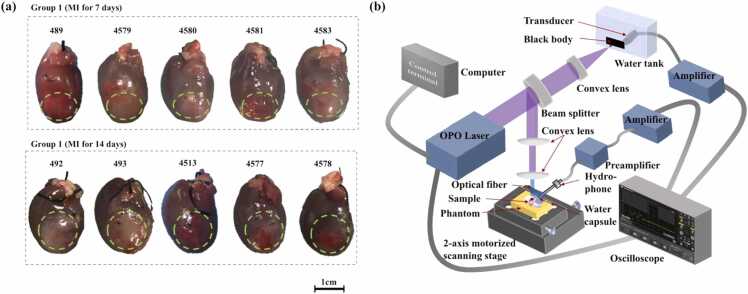
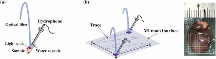
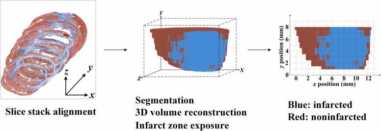
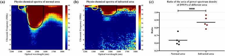
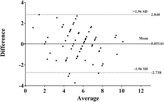
Myocardial infarction (MI) is a major cause of morbidity and mortality worldwide. Modern therapeutic strategies targeting the infarct border area have been shown to benefit overall cardiac function after MI. However, there is no non-invasive diagnostic technique to precisely demarcate the MI boundary till to now. In this study, the feasibility of demarcating the MI border using dual-wavelength photoacoustic spectral analysis (DWPASA) was investigated. To quantify specific molecular characteristics before and after MI, "the ratio of the areas of the power spectral densities ( )" was computed from the DWPASA results. Compared to the normal tissue, MI tissue was shown to contain more collagen, resulting in higher values ( < 0.001). Cross-sectional MI lengths and the MI area border demarcated in two dimensions by DWPASA were in substantial agreement with Masson staining (ICC = 0.76, < 0.001, IoU = 0.72). has been proved that can be used as an indicator of disease evolution to distinguish normal and pathological tissues. These findings indicate that the DWPASA method may offer a new diagnostic solution for determining MI borders.
心肌梗死(MI)是全球发病和死亡的主要原因。针对梗死边缘区域的现代治疗策略已被证明可改善心肌梗死后的整体心脏功能。然而,迄今为止,尚无精确划分心肌梗死边界的非侵入性诊断技术。在本研究中,研究了使用双波长光声光谱分析(DWPASA)划分心肌梗死边界的可行性。为了量化心肌梗死前后的特定分子特征,从DWPASA结果中计算“功率谱密度面积之比( )”。与正常组织相比,心肌梗死组织显示含有更多的胶原蛋白,导致更高的 值( <0.001)。DWPASA在二维上划分的心肌梗死横截面长度和心肌梗死面积边界与Masson染色结果基本一致(ICC = 0.76, <0.001,IoU = 0.72)。 已被证明可作为区分正常组织和病理组织的疾病演变指标。这些发现表明,DWPASA方法可能为确定心肌梗死边界提供一种新的诊断解决方案。