Department of Orthopaedic Surgery, Hebei Orthopaedic Clinical Research Center, The Third Hospital of Hebei Medical University, Shijiazhuang, China.
Key Laboratory of Biomechanics of Hebei Province, Orthopaedic Research Institute of Hebei Province, Hebei, China.
Orthop Surg. 2023 Dec;15(12):3279-3287. doi: 10.1111/os.13923. Epub 2023 Oct 19.
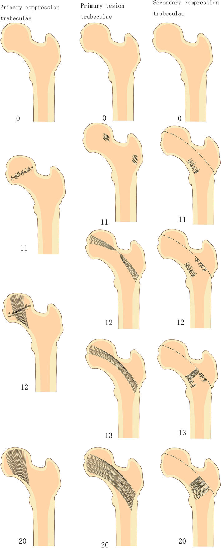
The Ward triangle is an important area used clinically to diagnose and assess osteoporosis and its fracture risk in the proximal femur. The main objective of this study was to investigate the rules of development and maturation of the trabeculae of Ward's triangle to provide a basis for the prevention and treatment proximal femur fracture.
From January 2018 to December 2019, individuals from 4 months to 19 years old who underwent hip growth and development assessments at the Third Hospital of Hebei Medical University were selected retrospectively. The outpatient electronic medical record system was used to collect information such as age, gender, imaging images, and clinical diagnosis. The development score and maturity characteristics of the trabecular bone were analyzed using hip radiograph data. Correlation analysis was performed to identify the relationship among age, neck-shaft angle and development and maturity score of the trabecular bone.
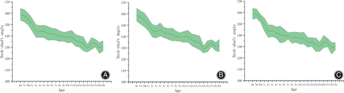
A total of 941 patients were enrolled in this study, including 539 males and 402 females. Primary compression trabeculae were all present at 1 year of age and matured at 7 years of age and older; primary tension trabeculae were all present at 4 years of age and matured at 18 years of age. Secondary compression trabeculae were present at 4 years of age and matured at 18 years of age. In addition, the neck-shaft angle progressively decreases from 4 months to 14 years of age but barely changes between 15 and 19 years of age.
In short, the development and maturation of the trabeculae in the ward' triangle followed a specific temporal pattern that was related to the neck-shaft angle. Therefore, these findings can help us understand structure and mechanical characteristics of proximal femoral trabeculae, and improve our understanding of the mechanism and treatment of proximal femoral fractures.
沃德尔三角(Ward triangle)是临床诊断和评估股骨近端骨质疏松症及其骨折风险的重要区域。本研究的主要目的是探讨沃德尔三角骨小梁的发育和成熟规律,为预防和治疗股骨近端骨折提供依据。
回顾性选取 2018 年 1 月至 2019 年 12 月在河北医科大学第三医院行髋关节生长发育评估的 4 月龄至 19 岁个体,应用门诊电子病历系统收集年龄、性别、影像学图像、临床诊断等信息,利用髋关节正位片数据,分析骨小梁的发育评分和成熟特征,采用相关性分析明确年龄、颈干角与骨小梁发育和成熟评分的关系。
本研究共纳入 941 例患者,其中男 539 例,女 402 例。1 岁时初级压应力骨小梁全部出现,7 岁以上成熟;4 岁时初级张应力骨小梁全部出现,18 岁时成熟;4 岁时次级压应力骨小梁出现,18 岁时成熟。颈干角从 4 月龄到 14 岁逐渐减小,而 15 岁到 19 岁之间几乎不变。
总之,沃德尔三角区骨小梁的发育和成熟遵循特定的时间模式,与颈干角有关。因此,这些发现有助于我们了解股骨近端骨小梁的结构和力学特征,提高我们对股骨近端骨折机制和治疗的认识。