• 文献检索
  • 文档翻译
  • 深度研究
  • 学术资讯
  • Suppr Zotero 插件Zotero 插件
  • 邀请有礼
  • 套餐&价格
  • 历史记录
应用&插件
Suppr Zotero 插件Zotero 插件浏览器插件Mac 客户端Windows 客户端微信小程序
定价
高级版会员购买积分包购买API积分包
服务
文献检索文档翻译深度研究API 文档MCP 服务
关于我们
关于 Suppr公司介绍联系我们用户协议隐私条款
关注我们

Suppr 超能文献

核心技术专利:CN118964589B侵权必究
粤ICP备2023148730 号-1Suppr @ 2026

文献检索

告别复杂PubMed语法,用中文像聊天一样搜索,搜遍4000万医学文献。AI智能推荐,让科研检索更轻松。

立即免费搜索

文件翻译

保留排版,准确专业,支持PDF/Word/PPT等文件格式,支持 12+语言互译。

免费翻译文档

深度研究

AI帮你快速写综述,25分钟生成高质量综述,智能提取关键信息,辅助科研写作。

立即免费体验

乳腺癌的合成磁共振成像:鉴别良恶性病变及预测免疫组化表达状态。

Synthetic MRI in breast cancer: differentiating benign from malignant lesions and predicting immunohistochemical expression status.

机构信息

Department of Medical Imaging, Shanxi Medical University, Taiyuan, Shanxi, China.

Department of Radiology, Cancer Hospital Chinese Academy of Medical Sciences, Shenzhen Center, Shenzhen, China.

出版信息

Sci Rep. 2023 Oct 20;13(1):17978. doi: 10.1038/s41598-023-45079-2.

DOI:10.1038/s41598-023-45079-2
PMID:37864025
原文链接:https://pmc.ncbi.nlm.nih.gov/articles/PMC10589282/
Abstract

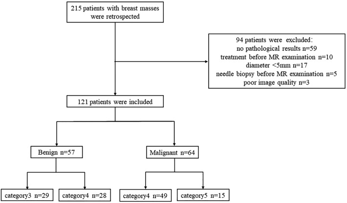
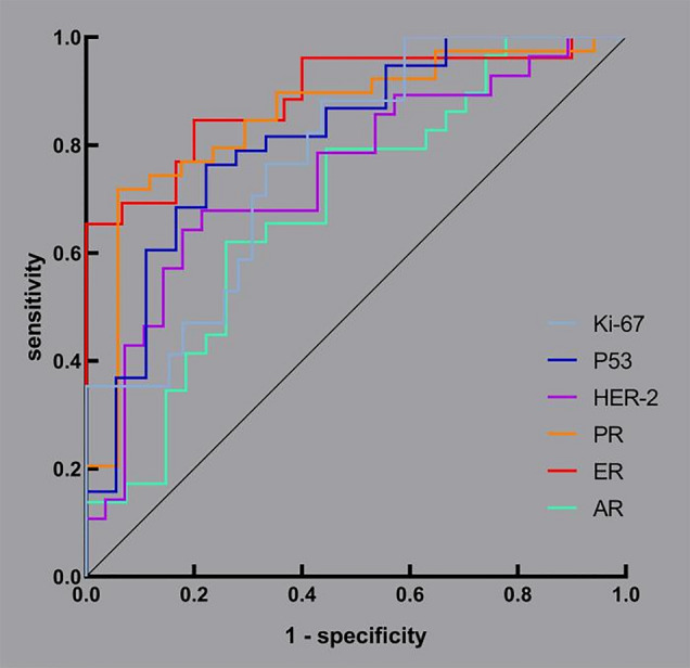
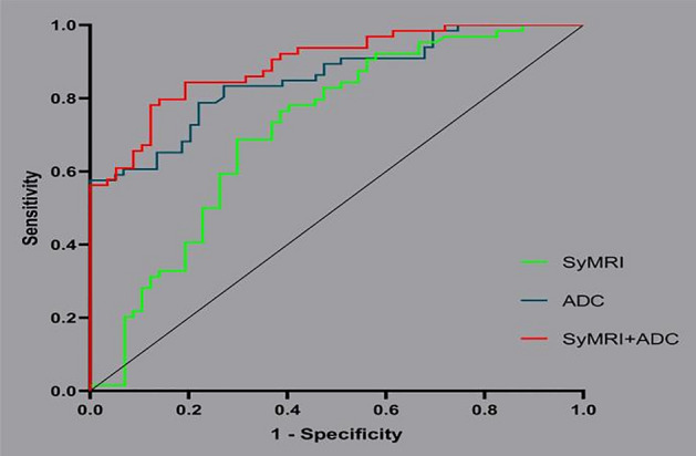
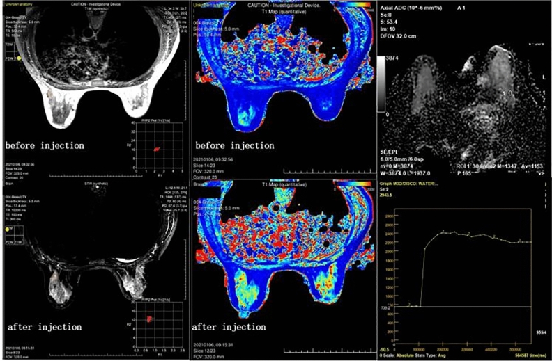
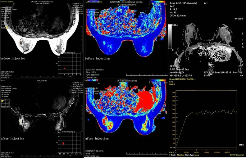
To evaluate and compare the performance of synthetic magnetic resonance imaging (SyMRI) in classifying benign and malignant breast lesions and predicting the expression status of immunohistochemistry (IHC) markers. We retrospectively analysed 121 patients with breast lesions who underwent dynamic contrast-enhanced magnetic resonance imaging (DCE-MRI) and SyMRI before surgery in our hospital. DCE-MRI was used to assess the lesions, and then regions of interest (ROIs) were outlined on SyMRI (before and after enhancement), and apparent diffusion coefficient (ADC) maps to obtain quantitative values. After being grouped according to benign and malignant status, the malignant lesions were divided into high and low expression groups according to the expression status of IHC markers. Logistic regression was used to analyse the differences in independent variables between groups. The performance of the modalities in classification and prediction was evaluated by receiver operating characteristic (ROC) curves. In total, 57 of 121 lesions were benign, the other 64 were malignant, and 56 malignant lesions performed immunohistochemical staining. Quantitative values from proton density-weighted imaging prior to an injection of the contrast agent (PD-Pre) and T2-weighted imaging (T2WI) after the injection (T2-Gd), as well as its standard deviation (SD of T2-Gd), were valuable SyMRI parameters for the classification of benign and malignant breast lesions, but the performance of SyMRI (area under the curve, AUC = 0.716) was not as good as that of ADC values (AUC = 0.853). However, ADC values could not predict the expression status of breast cancer markers, for which SyMRI had excellent performance. The AUCs of androgen receptor (AR), estrogen receptor (ER), progesterone receptor (PR), human epidermal growth factor receptor 2 (HER-2), p53 and Ki-67 were 0.687, 0.890, 0.852, 0.746, 0.813 and 0.774, respectively. SyMRI had certain value in distinguishing between benign and malignant breast lesions, and ADC values were still the ideal method. However, to predict the expression status of IHC markers, SyMRI had an incomparable value compared with ADC values.

摘要

为了评估和比较合成磁共振成像(SyMRI)在鉴别良恶性乳腺病变和预测免疫组织化学(IHC)标志物表达状态方面的性能。我们回顾性分析了 121 例在我院接受手术前动态对比增强磁共振成像(DCE-MRI)和 SyMRI 检查的乳腺病变患者。DCE-MRI 用于评估病变,然后在 SyMRI(增强前后)和表观扩散系数(ADC)图上勾画感兴趣区(ROI)以获得定量值。根据良恶性分组后,根据 IHC 标志物的表达状态,将恶性病变分为高表达和低表达组。采用逻辑回归分析组间独立变量的差异。通过受试者工作特征(ROC)曲线评估两种模式的分类和预测性能。121 个病灶中,57 个为良性,64 个为恶性,56 个恶性病灶行免疫组织化学染色。对比剂注射前质子密度加权成像(PD-Pre)和注射后 T2 加权成像(T2-Gd)的定量值及其标准差(T2-Gd 的 SD)是鉴别良恶性乳腺病变的有价值的 SyMRI 参数,但 SyMRI(曲线下面积,AUC=0.716)的性能不如 ADC 值(AUC=0.853)。然而,ADC 值不能预测乳腺癌标志物的表达状态,而 SyMRI 具有优异的性能。雄激素受体(AR)、雌激素受体(ER)、孕激素受体(PR)、人表皮生长因子受体 2(HER-2)、p53 和 Ki-67 的 AUC 分别为 0.687、0.890、0.852、0.746、0.813 和 0.774。SyMRI 在鉴别良恶性乳腺病变方面具有一定价值,ADC 值仍是理想方法。然而,预测 IHC 标志物的表达状态时,SyMRI 的价值无法与 ADC 值相比。

https://cdn.ncbi.nlm.nih.gov/pmc/blobs/f4ea/10589282/65dd2ccf1412/41598_2023_45079_Fig5_HTML.jpg
https://cdn.ncbi.nlm.nih.gov/pmc/blobs/f4ea/10589282/52167a5ea0f3/41598_2023_45079_Fig1_HTML.jpg
https://cdn.ncbi.nlm.nih.gov/pmc/blobs/f4ea/10589282/e0edd7c24f4a/41598_2023_45079_Fig2_HTML.jpg
https://cdn.ncbi.nlm.nih.gov/pmc/blobs/f4ea/10589282/2c6eec02bb1c/41598_2023_45079_Fig3_HTML.jpg
https://cdn.ncbi.nlm.nih.gov/pmc/blobs/f4ea/10589282/035715f95f9e/41598_2023_45079_Fig4_HTML.jpg
https://cdn.ncbi.nlm.nih.gov/pmc/blobs/f4ea/10589282/65dd2ccf1412/41598_2023_45079_Fig5_HTML.jpg

相似文献

1
Synthetic MRI in breast cancer: differentiating benign from malignant lesions and predicting immunohistochemical expression status.乳腺癌的合成磁共振成像:鉴别良恶性病变及预测免疫组化表达状态。
Sci Rep. 2023 Oct 20;13(1):17978. doi: 10.1038/s41598-023-45079-2.
2
[The value of synthetic MRI in differential diagnosis of benign and malignant breast lesions].[合成磁共振成像在乳腺良恶性病变鉴别诊断中的价值]
Zhonghua Zhong Liu Za Zhi. 2021 Aug 23;43(8):872-877. doi: 10.3760/cma.j.cn112152-20210322-00254.
3
[Differential diagnosis of benign and malignant breast lesions using quantitative synthetic magnetic resonance imaging].[利用定量合成磁共振成像对乳腺良恶性病变进行鉴别诊断]
Nan Fang Yi Ke Da Xue Xue Bao. 2022 Apr 20;42(4):457-462. doi: 10.12122/j.issn.1673-4254.2022.04.01.
4
Synthetic MRI and amide proton transfer-weighted MRI for differentiating between benign and malignant sinonasal lesions.合成磁共振成像和酰胺质子转移加权磁共振成像用于鉴别良恶性鼻窦病变。
Eur Radiol. 2024 Oct;34(10):6820-6830. doi: 10.1007/s00330-024-10696-6. Epub 2024 Mar 16.
5
The Role of Combined Diffusion-Weighted Imaging and Dynamic Contrast-Enhanced MRI for Differentiating Malignant From Benign Breast Lesions Presenting Washout Curve.联合扩散加权成像和动态对比增强 MRI 在鉴别呈洗脱曲线的良恶性乳腺病变中的作用。
Can Assoc Radiol J. 2021 Aug;72(3):460-469. doi: 10.1177/0846537120907098. Epub 2020 Mar 11.
6
Adding quantitative T1rho-weighted imaging to conventional MRI improves specificity and sensitivity for differentiating malignant from benign breast lesions.在传统磁共振成像(MRI)中加入定量T1rho加权成像可提高鉴别乳腺良恶性病变的特异性和敏感性。
Magn Reson Imaging. 2024 May;108:98-103. doi: 10.1016/j.mri.2024.02.005. Epub 2024 Feb 7.
7
Multiple parameters from ultrafast dynamic contrast-enhanced magnetic resonance imaging to discriminate between benign and malignant breast lesions: Comparison with apparent diffusion coefficient.从超快动态对比增强磁共振成像获取多个参数以鉴别乳腺良恶性病变:与表观扩散系数的比较。
Diagn Interv Imaging. 2023 Jun;104(6):275-283. doi: 10.1016/j.diii.2023.01.006. Epub 2023 Feb 2.
8
Investigation of synthetic MRI with quantitative parameters for discriminating axillary lymph nodes status in invasive breast cancer.采用合成 MRI 及定量参数对浸润性乳腺癌腋窝淋巴结状态进行鉴别诊断的研究。
Eur J Radiol. 2024 Jun;175:111452. doi: 10.1016/j.ejrad.2024.111452. Epub 2024 Apr 4.
9
Differentiating benign and malignant inflammatory breast lesions: Value of T2 weighted and diffusion weighted MR images.鉴别良性和恶性炎性乳腺病变:T2加权和扩散加权磁共振成像的价值
Magn Reson Imaging. 2018 Jul;50:38-44. doi: 10.1016/j.mri.2018.03.012. Epub 2018 Mar 12.
10
The diagnostic performance of quantitative mapping in breast cancer patients: a preliminary study using synthetic MRI.基于合成 MRI 的乳腺癌患者定量成像的诊断性能:初步研究
Cancer Imaging. 2020 Dec 14;20(1):88. doi: 10.1186/s40644-020-00365-4.

引用本文的文献

1
B1 corrected T1 mapping in the differentiation and prediction of breast cancer.B1校正T1映射在乳腺癌的鉴别诊断和预测中的应用
Sci Rep. 2025 Aug 21;15(1):30785. doi: 10.1038/s41598-025-15590-9.
2
Differentiation of breast cancer subtypes and correlation with biological status using functional magnetic resonance imaging: comparison with amide proton transfer-weighted imaging and diffusion-weighted imaging.利用功能磁共振成像对乳腺癌亚型进行鉴别及其与生物学状态的相关性:与酰胺质子转移加权成像和扩散加权成像的比较
Quant Imaging Med Surg. 2025 Jul 1;15(7):6102-6117. doi: 10.21037/qims-24-2174. Epub 2025 Jun 25.
3
Whole-tumor histogram analysis of synthetic MRI relaxation maps for nasopharyngeal carcinoma: correlations with histopathologic components.

本文引用的文献

1
Palbociclib for Residual High-Risk Invasive HR-Positive and HER2-Negative Early Breast Cancer-The Penelope-B Trial.帕博西尼用于残留高危侵袭性 HR 阳性和 HER2 阴性早期乳腺癌 - Penelope-B 试验。
J Clin Oncol. 2021 May 10;39(14):1518-1530. doi: 10.1200/JCO.20.03639. Epub 2021 Apr 1.
2
Diffusion-weighted Imaging Allows for Downgrading MR BI-RADS 4 Lesions in Contrast-enhanced MRI of the Breast to Avoid Unnecessary Biopsy.弥散加权成像可将乳腺增强 MRI 中 BI-RADS 4 级病变降级,避免不必要的活检。
Clin Cancer Res. 2021 Apr 1;27(7):1941-1948. doi: 10.1158/1078-0432.CCR-20-3037. Epub 2021 Jan 14.
3
The diagnostic performance of quantitative mapping in breast cancer patients: a preliminary study using synthetic MRI.
鼻咽癌合成磁共振成像弛豫图的全肿瘤直方图分析:与组织病理学成分的相关性
Jpn J Radiol. 2025 Jul 9. doi: 10.1007/s11604-025-01824-3.
4
Rapid multiparametric quantitative MRI for predicting the activity of thyroid-associated ophthalmopathy: combination with clinical characteristics.用于预测甲状腺相关性眼病活动度的快速多参数定量磁共振成像:与临床特征相结合
Eur Radiol. 2025 Jun 9. doi: 10.1007/s00330-025-11691-1.
5
FROP-1 peptide-conjugated ultrasmall superparamagnetic nanoparticles as a targeted T1-weighted MR contrast agent for breast cancer: in vitro study.FROP-1肽偶联的超小超顺磁性纳米颗粒作为乳腺癌的靶向T1加权磁共振造影剂:体外研究
BMC Biomed Eng. 2025 May 1;7(1):5. doi: 10.1186/s42490-025-00091-7.
6
Technical aspects and clinical applications of synthetic MRI: a scoping review.合成磁共振成像的技术层面与临床应用:一项范围综述
Diagnosis (Berl). 2025 Feb 7. doi: 10.1515/dx-2024-0168.
7
XGBoost Enhances the Performance of SAFE: A Novel Microwave Imaging System for Early Detection of Malignant Breast Cancer.XGBoost提升SAFE的性能:一种用于早期检测恶性乳腺癌的新型微波成像系统。
Cancers (Basel). 2025 Jan 10;17(2):214. doi: 10.3390/cancers17020214.
8
Multiparametric approach with synthetic MR imaging for diagnosing salivary gland lesions.多参数综合磁共振成像在唾液腺病变诊断中的应用。
Jpn J Radiol. 2024 Sep;42(9):983-992. doi: 10.1007/s11604-024-01578-4. Epub 2024 May 11.
基于合成 MRI 的乳腺癌患者定量成像的诊断性能:初步研究
Cancer Imaging. 2020 Dec 14;20(1):88. doi: 10.1186/s40644-020-00365-4.
4
The effect of gadolinium-based contrast agent administration on magnetic resonance fingerprinting-based T relaxometry in patients with prostate cancer.钆基造影剂给药对前列腺癌患者基于磁共振指纹技术的 T 弛豫时间测定的影响。
Sci Rep. 2020 Nov 24;10(1):20475. doi: 10.1038/s41598-020-77331-4.
5
Pretreatment structural and arterial spin labeling MRI is predictive for p53 mutation in high-grade gliomas.预处理结构和动脉自旋标记 MRI 可预测高级别胶质瘤中的 p53 突变。
Br J Radiol. 2020 Nov 1;93(1115):20200661. doi: 10.1259/bjr.20200661. Epub 2020 Sep 2.
6
Feasibility Study of Synthetic Diffusion-Weighted MRI in Patients with Breast Cancer in Comparison with Conventional Diffusion-Weighted MRI.合成扩散加权 MRI 与常规扩散加权 MRI 对比在乳腺癌患者中的可行性研究。
Korean J Radiol. 2020 Sep;21(9):1036-1044. doi: 10.3348/kjr.2019.0568.
7
Contrast-enhanced magnetic resonance (MR) T1 mapping with low-dose gadolinium-diethylenetriamine pentaacetic acid (Gd-DTPA) is promising in identifying clear cell renal cell carcinoma histopathological grade and differentiating fat-poor angiomyolipoma.使用低剂量钆喷酸葡胺(Gd-DTPA)的对比增强磁共振(MR)T1 映射在识别透明细胞肾细胞癌组织病理学分级和鉴别少脂性血管平滑肌脂肪瘤方面很有前景。
Quant Imaging Med Surg. 2020 May;10(5):988-998. doi: 10.21037/qims-19-723.
8
AR/ER Ratio Correlates with Expression of Proliferation Markers and with Distinct Subset of Breast Tumors.AR/ER 比值与增殖标志物的表达相关,并与特定的乳腺癌亚型相关。
Cells. 2020 Apr 24;9(4):1064. doi: 10.3390/cells9041064.
9
Utility of synthetic MRI in predicting the Ki-67 status of oestrogen receptor-positive breast cancer: a feasibility study.合成 MRI 在预测雌激素受体阳性乳腺癌 Ki-67 状态中的应用:一项可行性研究。
Clin Radiol. 2020 May;75(5):398.e1-398.e8. doi: 10.1016/j.crad.2019.12.021. Epub 2020 Feb 1.
10
Can apparent diffusion coefficient (ADC) distinguish breast cancer from benign breast findings? A meta-analysis based on 13 847 lesions.表观扩散系数(ADC)能否区分乳腺癌与良性乳腺病变?基于 13847 个病灶的荟萃分析。
BMC Cancer. 2019 Oct 15;19(1):955. doi: 10.1186/s12885-019-6201-4.