Jia Xu, Wu Jian, Chen Xiaohong, Hou Simeng, Li Yangyang, Zhao Ling, Zhu Yingting, Li Zhidong, Deng Caibin, Su Wenru, Zhuo Yehong
State Key Laboratory of Ophthalmology, Zhongshan Ophthalmic Center, Sun Yat-sen University, Guangdong Provincial Key Laboratory of Ophthalmology Visual Science, Guangzhou, Guangdong, China.
The Affiliated Hospital of Guizhou Medical University, Guiyang, Guizhou, China.
iScience. 2023 Sep 22;26(11):108024. doi: 10.1016/j.isci.2023.108024. eCollection 2023 Nov 17.
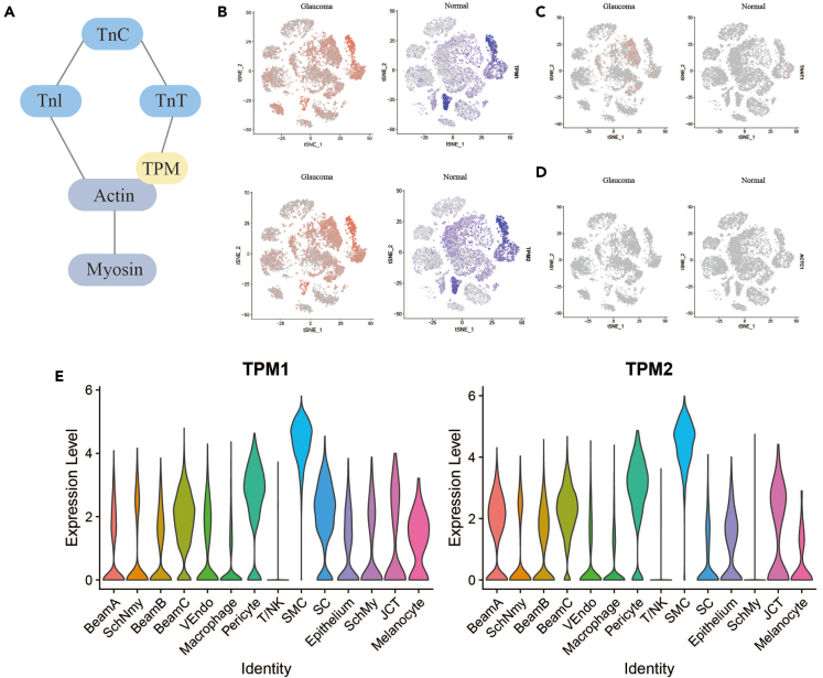
As the major channel of aqueous humor outflow, dysfunction of trabecular meshwork (TM) can lead to intraocular pressure elevating, which can trigger primary open-angle glaucoma (POAG). In this study, we use single-cell RNA sequencing (scRNA-seq) technique to build an atlas and further explore the spontaneous POAG and healthy macaques cellular heterogeneity associated with the dysfunction of TM contraction. We built the TM atlas, which identified 14 different cell types. In Beam A, Beam B, Beam C, and smooth muscle cell (SMC) cell types, we first found multiple genes associated with TM contraction (e.g., TPM1, ACTC1, TNNT1), determining their differential expression in the POAG and healthy groups. In addition, the microstructural alterations in TM of POAG non-human primates were observed, which was compact and collapsed. Thus, our study indicated that TPM1 may be a key target for regulating TM structure, contraction function, and resistance of aqueous humor outflow.
作为房水流出的主要通道,小梁网(TM)功能障碍可导致眼压升高,进而引发原发性开角型青光眼(POAG)。在本研究中,我们使用单细胞RNA测序(scRNA-seq)技术构建了一个图谱,并进一步探索与TM收缩功能障碍相关的自发性POAG和健康猕猴的细胞异质性。我们构建了TM图谱,鉴定出14种不同的细胞类型。在小梁束A、小梁束B、小梁束C和平滑肌细胞(SMC)细胞类型中,我们首次发现了多个与TM收缩相关的基因(如TPM1、ACTC1、TNNT1),并确定了它们在POAG组和健康组中的差异表达。此外,观察到POAG非人灵长类动物TM的微观结构改变,表现为紧密和塌陷。因此,我们的研究表明TPM1可能是调节TM结构、收缩功能和房水流出阻力的关键靶点。