Hu Yifan, Jiang Shanshan, Yu Xiaojin, Huang Sicong, Lan Ziting, Yu Yarong, Zhang Xiaohui, Chen Jin, Zhang Jiayin
Department of Radiology, Dongtai People's Hospital, Yancheng, China.
Department of Clinical and Technical Support, Philips Healthcare, Xi'an, China.
Quant Imaging Med Surg. 2023 Oct 1;13(10):6482-6492. doi: 10.21037/qims-23-233. Epub 2023 Aug 16.
Epicardial adipose tissue (EAT) is a key aspect in the investigation of cardiac pathophysiology. We sought to develop a deep learning (DL) model for fully automatic extraction and quantification of EAT through pulmonary computed tomography venography (PCTV) images.
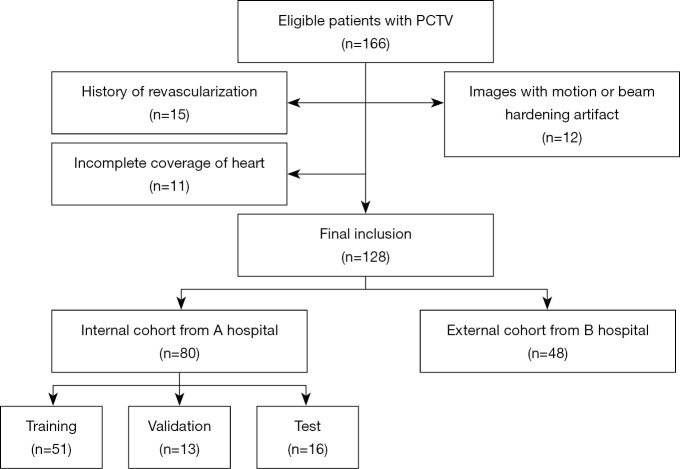
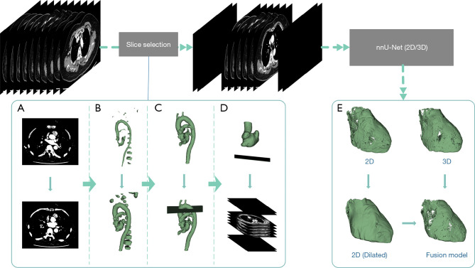
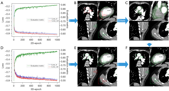
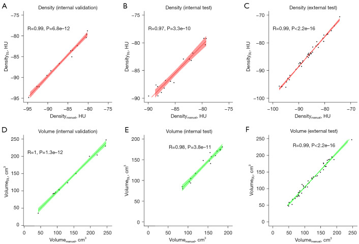
In this retrospective study, we included 128 patients with atrial fibrillation and PCTV from 2 hospitals. A DL model for automated EAT segmentation was developed from a training set of 51 patients and a validation set of 13 patients from hospital A. The algorithm was further validated using an internal test set of 16 patients from hospital A and an external test set of 48 patients from hospital B. The consistency and measurement agreement of EAT quantification were compared between the DL model and the conventional manual protocol using the Dice score coefficient (DSC), Hausdorff distance (HD95), Pearson correlation coefficient, and Bland-Altman plot.
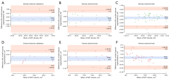
In the internal and external test set, automated segmentation with DL was successful in all cases. The total analysis time was shorter for DL than for manual reconstruction (5.43±2.52 106.20±15.90 min; P<0.001). The EAT segmented with the DL model had good consistency with manual segmentation (the DSC of the internal and external test sets were 0.92±0.02 and 0.88±0.03, respectively). The quantification of EAT evaluated with the 2 methods showed excellent correlation (all correlation coefficients >0.9; all P values <0.001) and minimal measurement difference.
The proposed DL model achieved fully automatic quantification of EAT from PCTV images. The yielded results were highly consistent with those of manual quantification.
心外膜脂肪组织(EAT)是心脏病理生理学研究的一个关键方面。我们试图开发一种深度学习(DL)模型,用于通过肺部计算机断层扫描静脉造影(PCTV)图像全自动提取和定量分析EAT。
在这项回顾性研究中,我们纳入了来自2家医院的128例房颤患者及PCTV图像。基于医院A的51例患者的训练集和13例患者的验证集开发了用于自动分割EAT的DL模型。该算法进一步使用医院A的16例患者的内部测试集和医院B的48例患者的外部测试集进行验证。使用Dice评分系数(DSC)、豪斯多夫距离(HD95)、皮尔逊相关系数和布兰德-奥特曼图,比较DL模型与传统手动方案之间EAT定量的一致性和测量一致性。
在内部和外部测试集中,DL自动分割在所有病例中均成功。DL的总分析时间比手动重建短(5.43±2.52对106.20±15.90分钟;P<0.001)。DL模型分割的EAT与手动分割具有良好的一致性(内部和外部测试集的DSC分别为0.92±0.02和0.88±0.03)。两种方法评估的EAT定量显示出极好的相关性(所有相关系数>0.9;所有P值<0.001)且测量差异最小。
所提出的DL模型实现了从PCTV图像中全自动定量分析EAT。所得结果与手动定量结果高度一致。