• 文献检索
  • 文档翻译
  • 深度研究
  • 学术资讯
  • Suppr Zotero 插件Zotero 插件
  • 邀请有礼
  • 套餐&价格
  • 历史记录
应用&插件
Suppr Zotero 插件Zotero 插件浏览器插件Mac 客户端Windows 客户端微信小程序
定价
高级版会员购买积分包购买API积分包
服务
文献检索文档翻译深度研究API 文档MCP 服务
关于我们
关于 Suppr公司介绍联系我们用户协议隐私条款
关注我们

Suppr 超能文献

核心技术专利:CN118964589B侵权必究
粤ICP备2023148730 号-1Suppr @ 2026

文献检索

告别复杂PubMed语法,用中文像聊天一样搜索,搜遍4000万医学文献。AI智能推荐,让科研检索更轻松。

立即免费搜索

文件翻译

保留排版,准确专业,支持PDF/Word/PPT等文件格式,支持 12+语言互译。

免费翻译文档

深度研究

AI帮你快速写综述,25分钟生成高质量综述,智能提取关键信息,辅助科研写作。

立即免费体验

用于在CT钙评分扫描中评估心外膜脂肪组织的深度学习分割和量化方法。

Deep learning segmentation and quantification method for assessing epicardial adipose tissue in CT calcium score scans.

作者信息

Hoori Ammar, Hu Tao, Lee Juhwan, Al-Kindi Sadeer, Rajagopalan Sanjay, Wilson David L

机构信息

Department of Biomedical Engineering, Case Western Reserve University, Cleveland, OH, 44106, USA.

Department of Cardiology, University Hospitals Cleveland Medical Center, Cleveland, OH, 44106, USA.

出版信息

Sci Rep. 2022 Feb 10;12(1):2276. doi: 10.1038/s41598-022-06351-z.

DOI:10.1038/s41598-022-06351-z
PMID:35145186
原文链接:https://pmc.ncbi.nlm.nih.gov/articles/PMC8831577/
Abstract

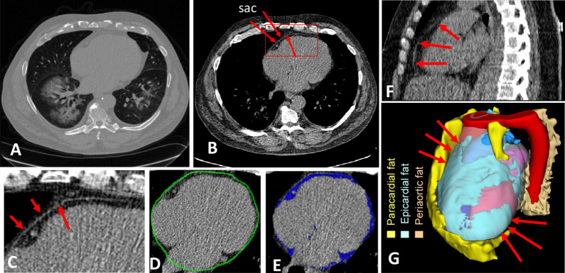
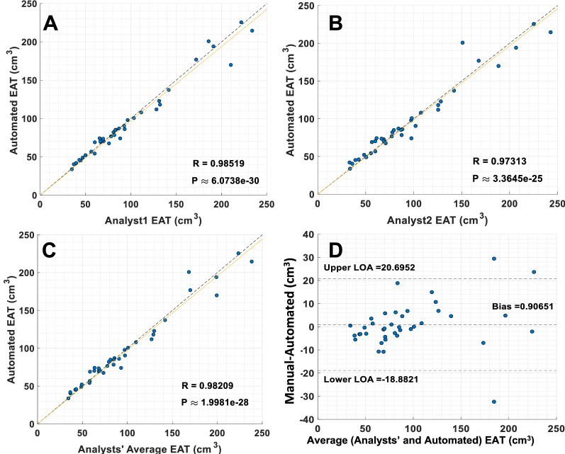
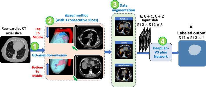
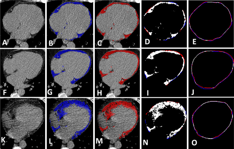
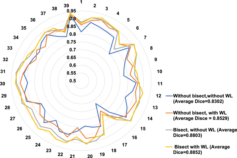
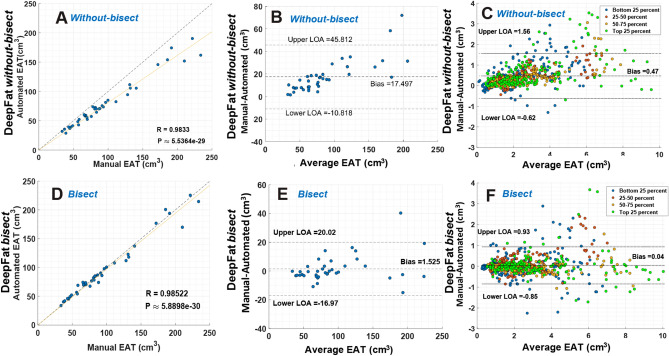
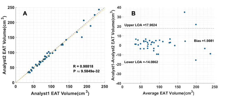
Epicardial adipose tissue volume (EAT) has been linked to coronary artery disease and the risk of major adverse cardiac events. As manual quantification of EAT is time-consuming, requires specialized training, and is prone to human error, we developed a deep learning method (DeepFat) for the automatic assessment of EAT on non-contrast low-dose CT calcium score images. Our DeepFat intuitively segmented the tissue enclosed by the pericardial sac on axial slices, using two preprocessing steps. First, we applied a HU-attention-window with a window/level 350/40-HU to draw attention to the sac and reduce numerical errors. Second, we applied a novel look ahead slab-of-slices with bisection ("bisect") in which we split the heart into halves and sequenced the lower half from bottom-to-middle and the upper half from top-to-middle, thereby presenting an always increasing curvature of the sac to the network. EAT volume was obtained by thresholding voxels within the sac in the fat window (- 190/- 30-HU). Compared to manual segmentation, our algorithm gave excellent results with volume Dice = 88.52% ± 3.3, slice Dice = 87.70% ± 7.5, EAT error = 0.5% ± 8.1, and R = 98.52% (p < 0.001). HU-attention-window and bisect improved Dice volume scores by 0.49% and 3.2% absolute, respectively. Variability between analysts was comparable to variability with DeepFat. Results compared favorably to those of previous publications.

摘要

心外膜脂肪组织体积(EAT)与冠状动脉疾病及主要不良心脏事件风险相关。由于手动定量EAT耗时、需要专业培训且容易出现人为误差,我们开发了一种深度学习方法(DeepFat),用于在非增强低剂量CT钙评分图像上自动评估EAT。我们的DeepFat使用两个预处理步骤,直观地在轴向切片上分割心包囊所包围的组织。首先,我们应用了一个窗宽/窗位为350/40 - HU的HU注意力窗口,以吸引对心包囊的关注并减少数值误差。其次,我们应用了一种新颖的带有二分法的前瞻性切片板(“二分法”),即将心脏分成两半,并对下半部分从底部到中部进行排序,对上半部分从顶部到中部进行排序,从而向网络呈现心包囊不断增加的曲率。通过对脂肪窗口(-190 / -30 - HU)内心包囊内的体素进行阈值处理来获得EAT体积。与手动分割相比,我们的算法取得了优异的结果,体积Dice系数 = 88.52% ± 3.3,切片Dice系数 = 87.70% ± 7.5,EAT误差 = 0.5% ± 8.1,相关系数R = 98.52%(p < 0.001)。HU注意力窗口和二分法分别使Dice体积分数绝对提高了0.49%和3.2%。分析人员之间的变异性与DeepFat的变异性相当。结果优于先前出版物中的结果。

https://cdn.ncbi.nlm.nih.gov/pmc/blobs/cb93/8831577/7603afa37c2f/41598_2022_6351_Fig7_HTML.jpg
https://cdn.ncbi.nlm.nih.gov/pmc/blobs/cb93/8831577/1ea594a72f66/41598_2022_6351_Fig1_HTML.jpg
https://cdn.ncbi.nlm.nih.gov/pmc/blobs/cb93/8831577/933a70497ce7/41598_2022_6351_Fig2_HTML.jpg
https://cdn.ncbi.nlm.nih.gov/pmc/blobs/cb93/8831577/f6e6f45f15aa/41598_2022_6351_Fig3_HTML.jpg
https://cdn.ncbi.nlm.nih.gov/pmc/blobs/cb93/8831577/26eed23a255f/41598_2022_6351_Fig4_HTML.jpg
https://cdn.ncbi.nlm.nih.gov/pmc/blobs/cb93/8831577/efe007f14940/41598_2022_6351_Fig5_HTML.jpg
https://cdn.ncbi.nlm.nih.gov/pmc/blobs/cb93/8831577/d1ae51745eb1/41598_2022_6351_Fig6_HTML.jpg
https://cdn.ncbi.nlm.nih.gov/pmc/blobs/cb93/8831577/7603afa37c2f/41598_2022_6351_Fig7_HTML.jpg

相似文献

1
Deep learning segmentation and quantification method for assessing epicardial adipose tissue in CT calcium score scans.用于在CT钙评分扫描中评估心外膜脂肪组织的深度学习分割和量化方法。
Sci Rep. 2022 Feb 10;12(1):2276. doi: 10.1038/s41598-022-06351-z.
2
Automatic Deep Learning Segmentation and Quantification of Epicardial Adipose Tissue in Non-Contrast Cardiac CT scans.自动深度学习分割和定量非对比心脏 CT 扫描中的心外膜脂肪组织。
Annu Int Conf IEEE Eng Med Biol Soc. 2021 Nov;2021:3938-3942. doi: 10.1109/EMBC46164.2021.9630953.
3
Automatic quantification of epicardial adipose tissue volume.自动量化心外膜脂肪组织体积。
Med Phys. 2021 Aug;48(8):4279-4290. doi: 10.1002/mp.15012. Epub 2021 Jun 29.
4
Deep Learning for Quantification of Epicardial and Thoracic Adipose Tissue From Non-Contrast CT.基于非对比 CT 的心脏外膜和胸腔脂肪组织定量的深度学习。
IEEE Trans Med Imaging. 2018 Aug;37(8):1835-1846. doi: 10.1109/TMI.2018.2804799. Epub 2018 Feb 9.
5
Deep Learning-Based Quantification of Epicardial Adipose Tissue Volume and Attenuation Predicts Major Adverse Cardiovascular Events in Asymptomatic Subjects.基于深度学习的心外膜脂肪组织体积和衰减定量预测无症状患者的主要不良心血管事件。
Circ Cardiovasc Imaging. 2020 Feb;13(2):e009829. doi: 10.1161/CIRCIMAGING.119.009829. Epub 2020 Feb 17.
6
Epicardial adipose tissue volume and annexin A2/fetuin-A signalling are linked to coronary calcification in advanced coronary artery disease: Computed tomography and proteomic biomarkers from the EPICHEART study.心外膜脂肪组织体积和膜联蛋白 A2/胎球蛋白-A 信号与晚期冠状动脉疾病中的冠状动脉钙化有关:来自 EPICHEART 研究的计算机断层扫描和蛋白质组学生物标志物。
Atherosclerosis. 2020 Jan;292:75-83. doi: 10.1016/j.atherosclerosis.2019.11.015. Epub 2019 Nov 15.
7
Segmentation and volume quantification of epicardial adipose tissue in computed tomography images.计算断层扫描图像中心外膜脂肪组织的分割和体积定量。
Med Phys. 2022 Oct;49(10):6477-6490. doi: 10.1002/mp.15965. Epub 2022 Sep 12.
8
A semi-automatic approach for epicardial adipose tissue segmentation and quantification on cardiac CT scans.一种用于心脏 CT 扫描上心外膜脂肪组织分割和定量的半自动方法。
Comput Biol Med. 2019 Nov;114:103424. doi: 10.1016/j.compbiomed.2019.103424. Epub 2019 Sep 5.
9
Statins Reduce Epicardial Adipose Tissue Attenuation Independent of Lipid Lowering: A Potential Pleiotropic Effect.他汀类药物降低心外膜脂肪组织衰减而不降低血脂:一种潜在的多效性效应。
J Am Heart Assoc. 2019 Jun 18;8(12):e013104. doi: 10.1161/JAHA.119.013104. Epub 2019 Jun 13.
10
Quantification of epicardial adipose tissue by cardiac CT: Influence of acquisition parameters and contrast enhancement.心脏 CT 测量心外膜脂肪组织:采集参数和对比增强的影响。
Eur J Radiol. 2019 Dec;121:108732. doi: 10.1016/j.ejrad.2019.108732. Epub 2019 Nov 1.

引用本文的文献

1
Radiomic features of peri-left atrial epicardial adipose tissue and atrial fibrillation recurrence after ablation.左心房周围心外膜脂肪组织的放射组学特征与消融术后房颤复发
Open Heart. 2025 Jul 8;12(2):e003364. doi: 10.1136/openhrt-2025-003364.
2
Opportunistic automated aortic valve calcification assessment in low-cost, screening CT calcium score exams.在低成本的筛查CT钙化积分检查中进行机会性自动主动脉瓣钙化评估。
Res Sq. 2025 Jun 2:rs.3.rs-6630820. doi: 10.21203/rs.3.rs-6630820/v1.
3
Detection of arterial remodeling using epicardial adipose tissue assessment from CT calcium scoring scan.

本文引用的文献

1
Epicardial fat attenuation, not volume, predicts obstructive coronary artery disease and high risk plaque features in patients with atypical chest pain.心外膜脂肪衰减而非体积预测非典型胸痛患者的阻塞性冠状动脉疾病和高危斑块特征。
Br J Radiol. 2020 Oct 1;93(1114):20200540. doi: 10.1259/bjr.20200540. Epub 2020 Aug 26.
2
Effects of Semaglutide Versus Dulaglutide on Epicardial Fat Thickness in Subjects with Type 2 Diabetes and Obesity.司美格鲁肽与度拉鲁肽对2型糖尿病合并肥胖症患者心外膜脂肪厚度的影响。
J Endocr Soc. 2020 Mar 13;4(4):bvz042. doi: 10.1210/jendso/bvz042. eCollection 2020 Apr 1.
3
Automatic segmentation and quantification of epicardial adipose tissue from coronary computed tomography angiography.
通过CT钙评分扫描的心外膜脂肪组织评估检测动脉重塑。
Front Cardiovasc Med. 2025 Mar 14;12:1543816. doi: 10.3389/fcvm.2025.1543816. eCollection 2025.
4
Evaluating Epicardial Fat Density Using ROI-Based Analysis: A Feasibility Study.使用基于感兴趣区域(ROI)分析评估心外膜脂肪密度:一项可行性研究。
J Cardiovasc Dev Dis. 2025 Feb 20;12(3):81. doi: 10.3390/jcdd12030081.
5
Prediction of obstructive coronary artery disease using coronary calcification and epicardial adipose tissue assessments from CT calcium scoring scans.利用CT钙评分扫描中的冠状动脉钙化和心外膜脂肪组织评估预测阻塞性冠状动脉疾病。
J Cardiovasc Comput Tomogr. 2025 Mar-Apr;19(2):224-231. doi: 10.1016/j.jcct.2025.01.007. Epub 2025 Feb 4.
6
Deep learning for opportunistic, end-to-end automated assessment of epicardial adipose tissue in pre-interventional, ECG-gated spiral computed tomography.深度学习用于在介入前心电图门控螺旋计算机断层扫描中对心外膜脂肪组织进行机会性、端到端自动评估。
Insights Imaging. 2024 Dec 19;15(1):301. doi: 10.1186/s13244-024-01875-6.
7
Leveraging calcium score CT radiomics for heart failure risk prediction.利用 CT 钙评分放射组学预测心力衰竭风险。
Sci Rep. 2024 Nov 6;14(1):26898. doi: 10.1038/s41598-024-77269-x.
8
An enhanced deep learning method for the quantification of epicardial adipose tissue.增强型深度学习方法用于心外膜脂肪组织定量。
Sci Rep. 2024 Oct 23;14(1):24947. doi: 10.1038/s41598-024-75659-9.
9
Artificial Intelligence Prediction of Cardiovascular Events Using Opportunistic Epicardial Adipose Tissue Assessments From Computed Tomography Calcium Score.利用计算机断层扫描钙评分进行机会性心外膜脂肪组织评估的心血管事件人工智能预测
JACC Adv. 2024 Aug 28;3(9):101188. doi: 10.1016/j.jacadv.2024.101188. eCollection 2024 Sep.
10
Deep learning analysis of epicardial adipose tissue to predict cardiovascular risk in heavy smokers.利用深度学习分析心外膜脂肪组织以预测重度吸烟者的心血管风险
Commun Med (Lond). 2024 Mar 13;4(1):44. doi: 10.1038/s43856-024-00475-1.
自动分割和定量冠状动脉计算机断层扫描血管造影中的心外膜脂肪组织。
Phys Med Biol. 2020 May 11;65(9):095012. doi: 10.1088/1361-6560/ab8077.
4
Fully Automated CT Quantification of Epicardial Adipose Tissue by Deep Learning: A Multicenter Study.深度学习实现的心脏外膜脂肪组织全自动化CT定量分析:一项多中心研究
Radiol Artif Intell. 2019 Nov 27;1(6):e190045. doi: 10.1148/ryai.2019190045.
5
Generalizing Deep Learning for Medical Image Segmentation to Unseen Domains via Deep Stacked Transformation.通过深度堆叠变换将深度学习用于医学图像分割推广到未见领域。
IEEE Trans Med Imaging. 2020 Jul;39(7):2531-2540. doi: 10.1109/TMI.2020.2973595. Epub 2020 Feb 12.
6
Fast fully automatic heart fat segmentation in computed tomography datasets.快速全自动心脏脂肪分割在计算机断层扫描数据集。
Comput Med Imaging Graph. 2020 Mar;80:101674. doi: 10.1016/j.compmedimag.2019.101674. Epub 2019 Dec 6.
7
Association of epicardial adipose tissue attenuation with coronary atherosclerosis in patients with a high risk of coronary artery disease.心外膜脂肪组织衰减与冠心病高危患者冠状动脉粥样硬化的关系。
Atherosclerosis. 2019 May;284:230-236. doi: 10.1016/j.atherosclerosis.2019.01.033. Epub 2019 Feb 6.
8
Epicardial Fat Volume Improves the Prediction of Obstructive Coronary Artery Disease Above Traditional Risk Factors and Coronary Calcium Score.心外膜脂肪体积可提高传统危险因素和冠状动脉钙评分对阻塞性冠状动脉疾病的预测价值。
Circ Cardiovasc Imaging. 2019 Jan;12(1):e008002. doi: 10.1161/CIRCIMAGING.118.008002.
9
Deep Learning for Quantification of Epicardial and Thoracic Adipose Tissue From Non-Contrast CT.基于非对比 CT 的心脏外膜和胸腔脂肪组织定量的深度学习。
IEEE Trans Med Imaging. 2018 Aug;37(8):1835-1846. doi: 10.1109/TMI.2018.2804799. Epub 2018 Feb 9.
10
Epicardial adipose tissue volume assessed by computed tomography and coronary artery disease: a systematic review and meta-analysis.计算机断层扫描评估的心外膜脂肪组织体积与冠状动脉疾病:系统评价和荟萃分析。
Eur Heart J Cardiovasc Imaging. 2018 May 1;19(5):490-497. doi: 10.1093/ehjci/jex314.