Wang Aijie, Li Wanjiang, Huang Wenyu, Luo Mao, Xiao Wendan, Qin Chaoyi, Dong Shushan, Liu Haiwei, Li Zhenlin, Diao Kaiyue
Department of Radiology, West China Hospital of Sichuan University, Chengdu, China.
West China School of Medicine, West China Hospital of Sichuan University, Chengdu, China.
Quant Imaging Med Surg. 2023 Oct 1;13(10):6456-6467. doi: 10.21037/qims-23-101. Epub 2023 Sep 18.
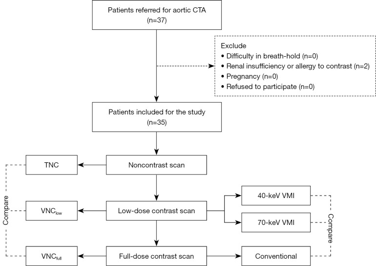
Computed tomography angiography (CTA) is the recommended diagnostic and follow-up imaging modality for acute aortic dissection (AD). However, the high-contrast medium burden associated with repeated CT aortography follow-ups remains a significant concern. This prospective study aimed to assess whether an ultra-low contrast dose (75% cutoff) aortic CTA protocol on dual-layer spectral CT could achieve comparable image quality with the full dose protocol. We also investigated the image quality of the virtual noncontrast (VNC) images derived from the ultra-low dose protocol.
This study included 37 consecutive patients who were referred to aortic CTA from May 2022 to August 2022. The enrolled patients underwent full-dose contrast CTA and ultra-low dose (reduced to 25% of conventional) contrast CTA on dual-layer spectral CT in 1 day. Virtual monochromatic images (VMIs) were reconstructed with 40 and 70 keV. The VNC images were reconstructed for both protocols. Objective image quality evaluation, recorded as signal-to-noise ratios (SNRs) and contrast-to-noise ratios (CNRs), was compared between the groups using 1-way analysis of variance and post hoc analysis with Bonferroni correction. Subjective image quality was also compared between the groups. Finally, VNC images derived from the low-dose (VNC) and full-dose (VNC) protocols were compared to the true noncontrast (TNC) images.
Neither CNR nor SNR was lower for the 40-keV images reconstructed from the ultra-low dose group compared to the conventional images. Both were significantly higher than those of the 70-keV images. Regarding subjective image quality, vessel enhancement was not significantly different between the 40-keV VMI and full-dose images [ascending aorta (AAO): 4.37±0.46 4.57±0.48, P=0.096; brachiocephalic arteries: 4.34±0.45 4.51±0.49, P=0.152; abdominal aortic side branch: 4.42±0.48 4.51±0.49, P=0.480]. The VNC images were similar to the TNC images but significantly different from the VNC images (P<0.001).
Ultra-low contrast aortic CTA with a 75%-reduced iodine dose using dual-layer spectral CT and the derived VNC achieved image quality comparable to that of conventional CTA and TNC images.
计算机断层血管造影(CTA)是急性主动脉夹层(AD)推荐的诊断和随访成像方式。然而,重复进行CT主动脉造影随访所带来的高对比剂负荷仍是一个重大问题。这项前瞻性研究旨在评估双层光谱CT上的超低对比剂剂量(75%剂量截断)主动脉CTA方案是否能获得与全剂量方案相当的图像质量。我们还研究了源自超低剂量方案的虚拟平扫(VNC)图像的质量。
本研究纳入了2022年5月至2022年8月期间连续37例接受主动脉CTA检查的患者。入选患者在1天内于双层光谱CT上接受全剂量对比剂CTA和超低剂量(降至传统剂量的25%)对比剂CTA检查。使用40 keV和70 keV重建虚拟单色图像(VMI)。两种方案均重建VNC图像。采用单因素方差分析和Bonferroni校正的事后分析比较两组之间以信噪比(SNR)和对比噪声比(CNR)记录的客观图像质量评估结果。两组之间也比较主观图像质量。最后,将源自低剂量(VNC)和全剂量(VNC)方案的VNC图像与真实平扫(TNC)图像进行比较。
与传统图像相比,超低剂量组重建的40 keV图像的CNR和SNR均未降低。两者均显著高于70 keV图像。关于主观图像质量,40 keV VMI与全剂量图像之间的血管强化无显著差异[升主动脉(AAO):4.37±0.46对4.57±0.48,P = 0.096;头臂动脉:4.3 ± 0.45对4.51±0.49,P = 0.152;腹主动脉分支:4.42±0.48对4.51±0.49,P = 0.480]。VNC图像与TNC图像相似,但与VNC图像有显著差异(P < 0.001)。
使用双层光谱CT且碘剂量降低75%的超低对比剂主动脉CTA及其衍生的VNC图像质量与传统CTA和TNC图像相当。