Tanoue Shota, Nakaura Takeshi, Nagayama Yasunori, Uetani Hiroyuki, Ikeda Osamu, Yamashita Yasuyuki
Department of Diagnostic Radiology, Graduate School of Medical Sciences, Kumamoto University, Kumamoto, Japan.
Korean J Radiol. 2021 Jun;22(6):951-958. doi: 10.3348/kjr.2020.0677. Epub 2021 Jan 29.
To evaluate the usefulness of virtual monochromatic images (VMIs) obtained using dual-layer dual-energy CT (DL-DECT) for evaluating brain tumors.
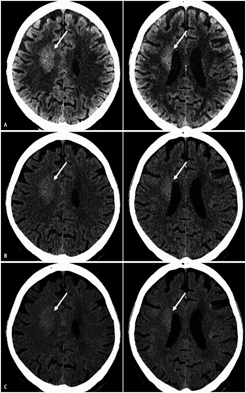
This retrospective study included 32 patients with brain tumors who had undergone non-contrast head CT using DL-DECT. Among them, 15 had glioblastoma (GBM), 7 had malignant lymphoma, 5 had high-grade glioma other than GBM, 3 had low-grade glioma, and 2 had metastatic tumors. Conventional polychromatic images and VMIs (40-200 keV at 10 keV intervals) were generated. We compared CT attenuation, image noise, contrast, and contrast-to-noise ratio (CNR) between tumor and white matter (WM) or grey matter (GM) between VMIs showing the highest CNR (optimized VMI) and conventional CT images using the paired t test. Two radiologists subjectively assessed the contrast, margin, noise, artifact, and diagnostic confidence of optimized VMIs and conventional images on a 4-point scale.
The image noise of VMIs at all energy levels tested was significantly lower than that of conventional CT images ( < 0.05). The 40-keV VMIs yielded the best CNR. Furthermore, both contrast and CNR between the tumor and WM were significantly higher in the 40 keV images than in the conventional CT images ( < 0.001); however, the contrast and CNR between tumor and GM were not significantly different ( = 0.47 and = 0.31, respectively). The subjective scores assigned to contrast, margin, and diagnostic confidence were significantly higher for 40 keV images than for conventional CT images ( < 0.01).
In head CT for patients with brain tumors, compared with conventional CT images, 40 keV VMIs from DL-DECT yielded superior tumor contrast and diagnostic confidence, especially for brain tumors located in the WM.
评估使用双层双能量CT(DL-DECT)获得的虚拟单色图像(VMI)对脑肿瘤评估的有效性。
这项回顾性研究纳入了32例接受DL-DECT非增强头部CT检查的脑肿瘤患者。其中,15例为胶质母细胞瘤(GBM),7例为恶性淋巴瘤,5例为非GBM的高级别胶质瘤,3例为低级别胶质瘤,2例为转移瘤。生成了常规多色图像和VMI(40 - 200 keV,间隔10 keV)。我们使用配对t检验比较了显示最高对比噪声比(CNR)的VMI(优化后的VMI)与常规CT图像之间肿瘤与白质(WM)或灰质(GM)的CT衰减、图像噪声、对比度和对比噪声比(CNR)。两名放射科医生以4分制主观评估优化后的VMI和常规图像的对比度、边缘、噪声、伪影和诊断信心。
所有测试能量水平的VMI图像噪声均显著低于常规CT图像(<0.05)。40 keV的VMI产生了最佳的CNR。此外,40 keV图像中肿瘤与WM之间的对比度和CNR均显著高于常规CT图像(<0.001);然而,肿瘤与GM之间的对比度和CNR无显著差异(分别为=0.47和=0.31)。40 keV图像在对比度、边缘和诊断信心方面的主观评分显著高于常规CT图像(<0.01)。
在脑肿瘤患者的头部CT检查中,与常规CT图像相比,DL-DECT的40 keV VMI具有更高的肿瘤对比度和诊断信心,特别是对于位于WM的脑肿瘤。