Laboratory of Pathophysiology, University of Antwerp, Universiteitsplein 1, 2610, Antwerp, Belgium.
Receptor Biology Lab, Department of Biomedical Science, University of Antwerp, Antwerp, Belgium.
Sci Rep. 2023 Oct 23;13(1):18119. doi: 10.1038/s41598-023-43567-z.
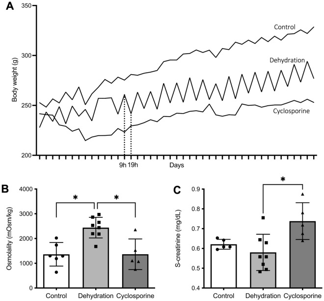
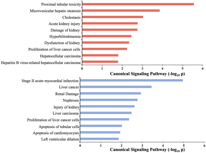
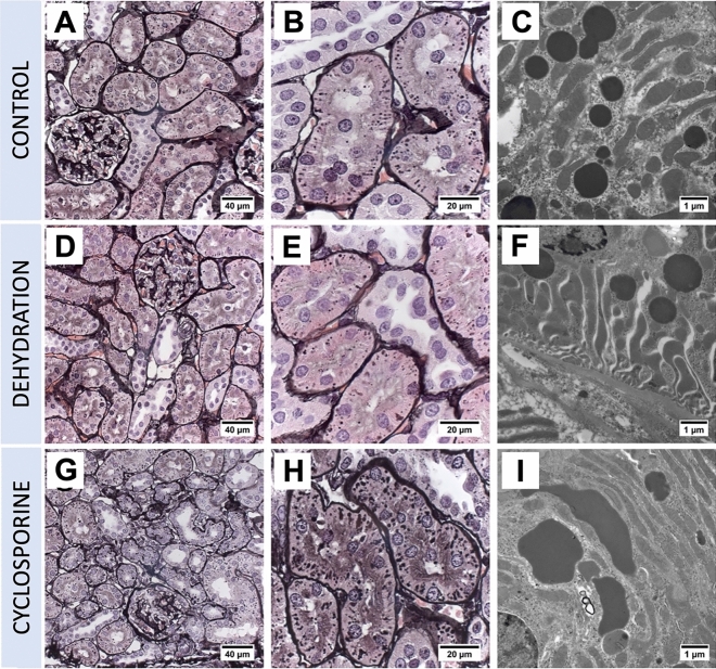
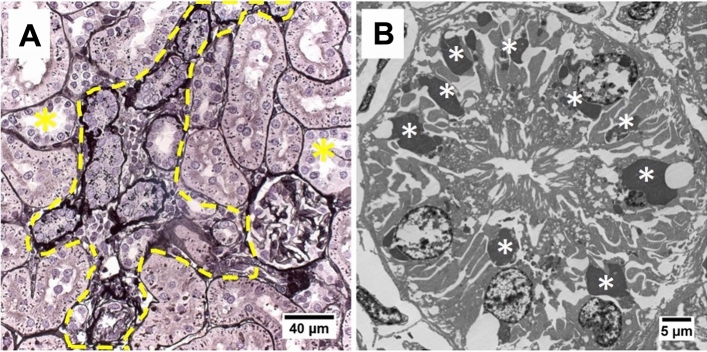
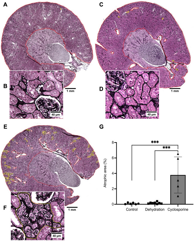
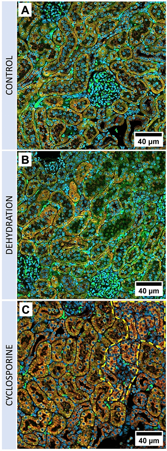
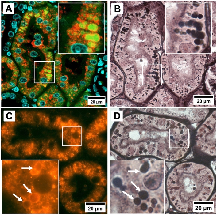
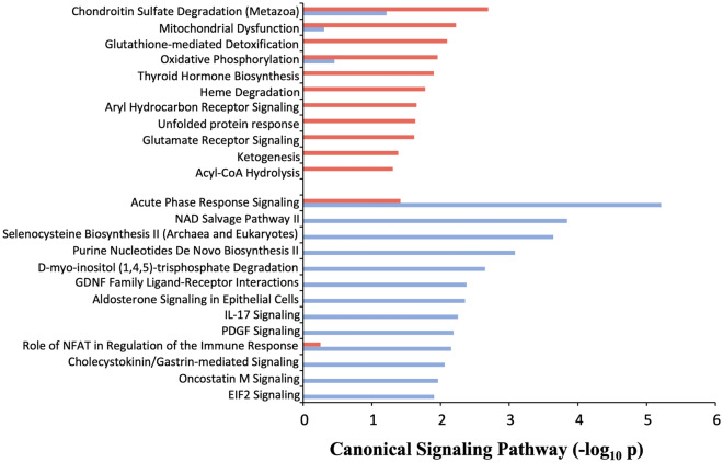
CINAC-patients present renal proximal tubular cell lysosomal lesions which are also observed in patients experiencing calcineurin inhibitor (CNI) nephrotoxicity, suggesting that CINAC is a toxin-induced nephropathy. An alternative hypothesis advocates chronic dehydration as a major etiological factor for CINAC. Here, we evaluated histological and molecular changes in dehydrated versus toxin exposed rats. Wistar rats were divided in 3 groups. Group 1 (n = 6) had free access to drinking water (control group). Group 2 (n = 8) was water deprived for 10 h per 24 h, 5 days/week and placed in an incubator (37 °C) for 30 min/h during water deprivation. Group 3 (n = 8) underwent daily oral gavage with cyclosporine (40 mg/kg body weight). After 28 days, renal function, histopathology and proteomic signatures were analysed. Cyclosporine-treated rats developed focal regions of atrophic proximal tubules with associated tubulo-interstitial fibrosis. PASM staining revealed enlarged argyrophilic granules in affected proximal tubules, identified as lysosomes by immunofluorescent staining. Electron microscopy confirmed the enlarged and dysmorphic phenotype of the lysosomes. Overall, these kidney lesions resemble those that have been previously documented in farmers with CINAC. Dehydration resulted in none of the above histopathological features. Proteomic analysis revealed that dehydration and cyclosporine both induce injury pathways, yet of a clear distinct nature with a signature of toxicity only for the cyclosporine group. In conclusion, both cyclosporine and dehydration are injurious to the kidney. However, dehydration alone does not result in kidney histopathology as observed in CINAC patients, whereas cyclosporine administration does. The histopathological analogy between CINAC and calcineurin inhibitor nephrotoxicity in rats and humans supports the involvement of an as-yet-unidentified environmental toxin in CINAC etiology.
CINAC 患者表现出肾近端管状细胞溶酶体损伤,这也见于经历钙调磷酸酶抑制剂 (CNI) 肾毒性的患者,表明 CINAC 是一种毒素诱导的肾病。另一种假说主张慢性脱水是 CINAC 的主要病因因素。在这里,我们评估了脱水与毒素暴露的大鼠的组织学和分子变化。Wistar 大鼠分为 3 组。第 1 组(n=6)自由饮用饮用水(对照组)。第 2 组(n=8)每 24 小时饮水 10 小时,每周 5 天,并在脱水期间每 30 分钟置于孵育箱(37°C)中 30 分钟。第 3 组(n=8)每天口服环孢素(40mg/kg 体重)。28 天后,分析肾功能、组织病理学和蛋白质组学特征。环孢素治疗的大鼠出现局灶性萎缩近端肾小管区域,并伴有肾小管间质纤维化。PASM 染色显示受影响的近端肾小管中增大的嗜银颗粒,通过免疫荧光染色鉴定为溶酶体。电子显微镜证实了溶酶体的增大和畸形表型。总的来说,这些肾脏病变与以前在患有 CINAC 的农民中记录的病变相似。脱水没有导致上述任何组织病理学特征。蛋白质组学分析表明,脱水和环孢素都诱导损伤途径,但性质明显不同,只有环孢素组具有毒性特征。总之,环孢素和脱水都会对肾脏造成损伤。然而,单独脱水不会导致 CINAC 患者观察到的肾脏组织病理学,而环孢素给药会导致。CINAC 与大鼠和人类钙调磷酸酶抑制剂肾毒性的组织病理学相似性支持了一种尚未确定的环境毒素在 CINAC 病因中的参与。