Department of Mechanical Engineering, Boston University, 110 Cummington Mall, Boston, MA, 02215, USA.
Department of Biomedical Engineering, Boston University, Boston, MA, 02215, USA.
Alzheimers Res Ther. 2023 Oct 27;15(1):185. doi: 10.1186/s13195-023-01331-5.
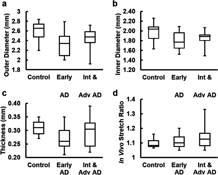
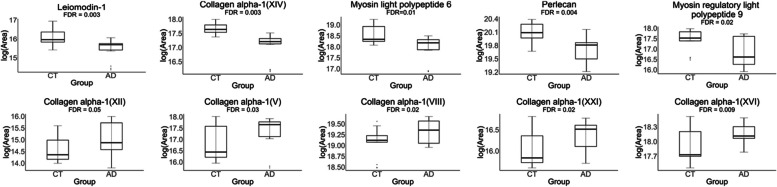
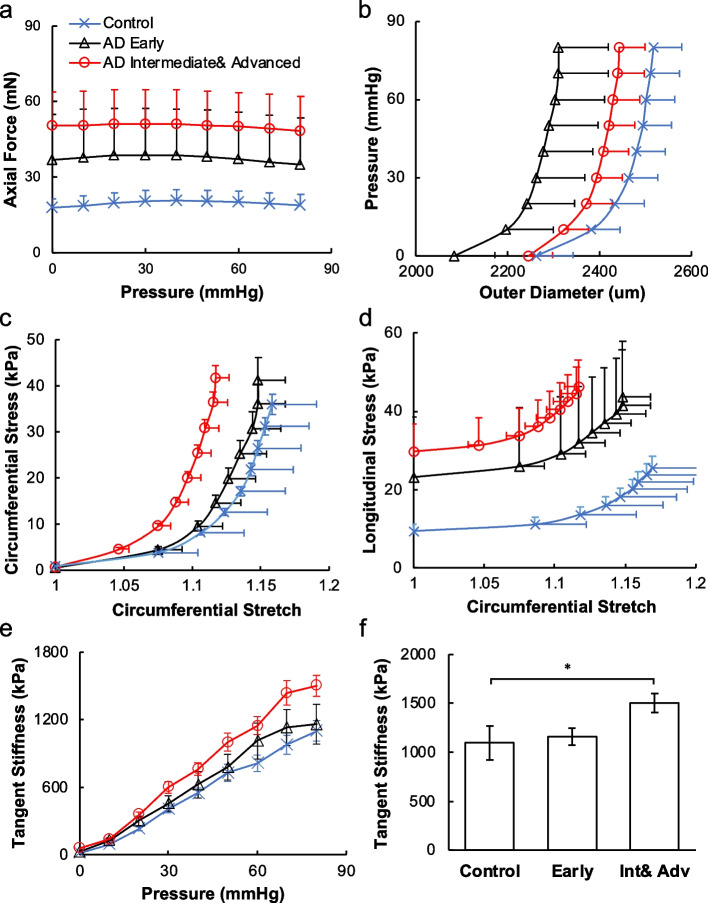
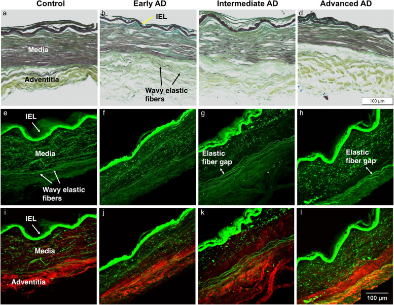
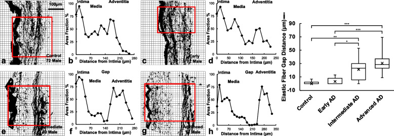
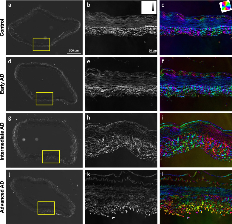
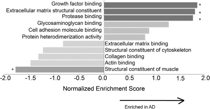
Alzheimer's disease (AD) is a neurodegenerative disease and the main cause for dementia. The irreversible neurodegeneration leads to a gradual loss of brain function characterized predominantly by memory loss. Cerebrovascular changes are common neuropathologic findings in aged subjects with dementia. Cerebrovascular integrity is critical for proper metabolism and perfusion of the brain, as cerebrovascular remodeling may render the brain more susceptible to pulse pressure and may be associated with poorer cognitive performance and greater risk of cerebrovascular events. The objective of this study is to provide understanding of cerebrovascular remodeling with AD progression. Anterior cerebral arteries (ACAs) from a total of 19 brain donor participants from controls and pathologically diagnosed AD groups (early-Braak stages I-II; intermediate-Braak stages III-IV; and advanced-Braak stages V-VI) were included in this study. Mechanical testing, histology, advanced optical imaging, and mass spectrometry were performed to study the progressive structural and functional changes of ACAs with AD progression. Biaxial extension-inflation tests showed that ACAs became progressively less compliant, and the longitudinal stress in the intermediate and advanced AD groups was significantly higher than that from the control group. With pathological AD development, the inner and outer diameters of the ACAs remained almost unchanged; however, histology study revealed progressive smooth muscle cell atrophy and loss of elastic fibers which led to compromised structural integrity of the arterial wall. Multiphoton imaging demonstrated elastin degradation at the media-adventitia interface, which led to the formation of an empty band of 21.0 ± 15.4 μm and 32.8 ± 9.24 μm in width for the intermediate and advanced AD groups, respectively. Furthermore, quantitative birefringence microscopy showed disorganized adventitial collagen with AD development. Mass spectrometry analysis provided further evidence of altered collagen content and other extracellular matrix (ECM) molecule and smooth muscle cell changes that were consistent with the mechanical and structural alterations. Collectively, our study provides understanding of the mechanical and structural cerebrovascular deterioration in cerebral arteries with AD, which may be related to neurodegenration and pathology in the brain.
阿尔茨海默病(AD)是一种神经退行性疾病,也是痴呆的主要病因。不可逆转的神经退行性变导致脑功能逐渐丧失,主要表现为记忆力丧失。脑血管变化是老年痴呆患者常见的神经病理学发现。脑血管完整性对于大脑的正常代谢和灌注至关重要,因为脑血管重塑可能使大脑更容易受到脉压的影响,并可能与认知表现较差和发生脑血管事件的风险增加有关。本研究的目的是了解 AD 进展过程中的脑血管重塑。本研究共纳入了 19 名脑捐献者的大脑前动脉(ACAs),这些捐献者来自对照组和病理诊断为 AD 的组(早期 Braak 阶段 I-II;中期 Braak 阶段 III-IV;晚期 Braak 阶段 V-VI)。进行了力学测试、组织学、高级光学成像和质谱分析,以研究随着 AD 进展,ACAs 的进行性结构和功能变化。双轴拉伸-膨胀测试表明,ACAs 的顺应性逐渐降低,中期和晚期 AD 组的纵向应力明显高于对照组。随着 AD 病理的发展,ACAs 的内、外径几乎不变;然而,组织学研究显示平滑肌细胞逐渐萎缩和弹性纤维丢失,导致动脉壁的结构完整性受损。多光子成像显示中膜-外膜交界处的弹性蛋白降解,导致中间和晚期 AD 组分别形成 21.0±15.4μm 和 32.8±9.24μm 宽的空带。此外,定量双折射显微镜显示 AD 发展过程中 Adventitial 胶原排列紊乱。质谱分析提供了进一步的证据,表明胶原含量和其他细胞外基质(ECM)分子以及平滑肌细胞的变化与力学和结构改变一致。总之,我们的研究提供了对 AD 大脑中脑血管力学和结构恶化的认识,这可能与大脑中的神经退行性变和病理学有关。