• 文献检索
  • 文档翻译
  • 深度研究
  • 学术资讯
  • Suppr Zotero 插件Zotero 插件
  • 邀请有礼
  • 套餐&价格
  • 历史记录
应用&插件
Suppr Zotero 插件Zotero 插件浏览器插件Mac 客户端Windows 客户端微信小程序
定价
高级版会员购买积分包购买API积分包
服务
文献检索文档翻译深度研究API 文档MCP 服务
关于我们
关于 Suppr公司介绍联系我们用户协议隐私条款
关注我们

Suppr 超能文献

核心技术专利:CN118964589B侵权必究
粤ICP备2023148730 号-1Suppr @ 2026

文献检索

告别复杂PubMed语法,用中文像聊天一样搜索,搜遍4000万医学文献。AI智能推荐,让科研检索更轻松。

立即免费搜索

文件翻译

保留排版,准确专业,支持PDF/Word/PPT等文件格式,支持 12+语言互译。

免费翻译文档

深度研究

AI帮你快速写综述,25分钟生成高质量综述,智能提取关键信息,辅助科研写作。

立即免费体验

2D 还是非 2D? 角膜巩膜壳胶原纤维的深度倾斜测绘。

2D or not 2D? Mapping the in-depth inclination of the collagen fibers of the corneoscleral shell.

机构信息

Department of Ophthalmology, University of Pittsburgh, Pittsburgh, PA, USA; Department of Bioengineering, University of Pittsburgh, Pittsburgh, PA, USA.

Department of Ophthalmology, University of Pittsburgh, Pittsburgh, PA, USA.

出版信息

Exp Eye Res. 2023 Dec;237:109701. doi: 10.1016/j.exer.2023.109701. Epub 2023 Oct 26.

DOI:10.1016/j.exer.2023.109701
PMID:37898229
原文链接:https://pmc.ncbi.nlm.nih.gov/articles/PMC10872428/
Abstract

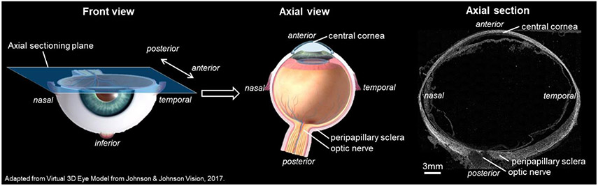
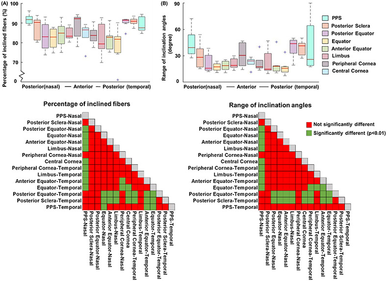
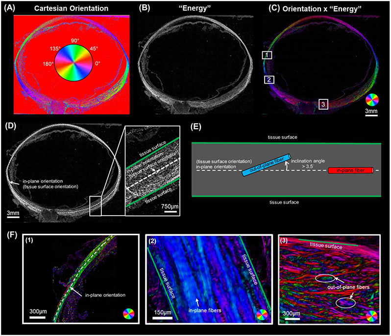
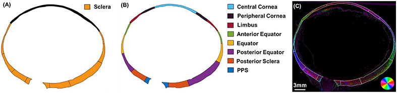
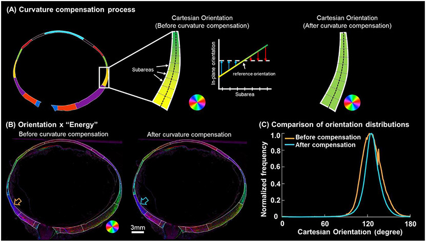
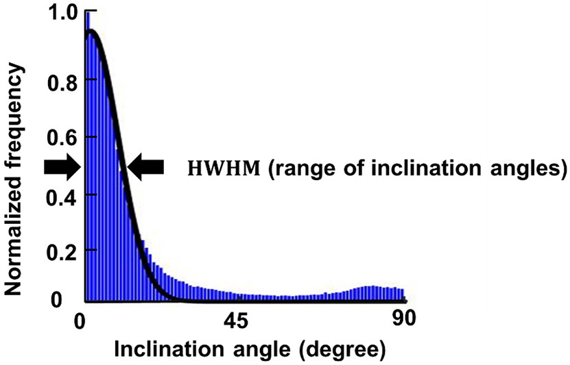
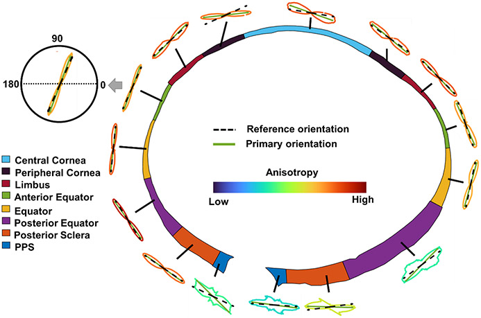
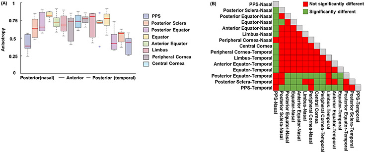
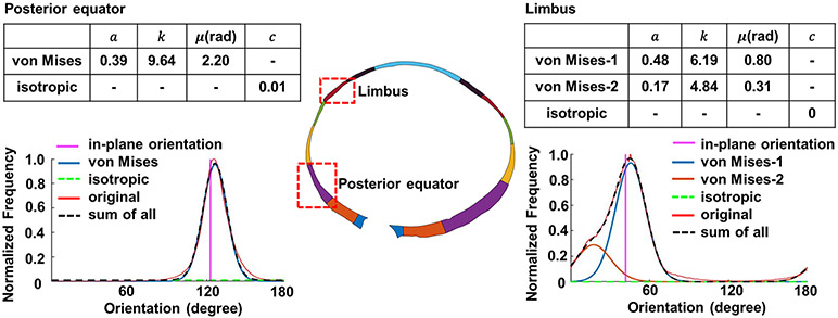
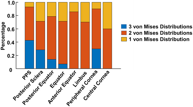
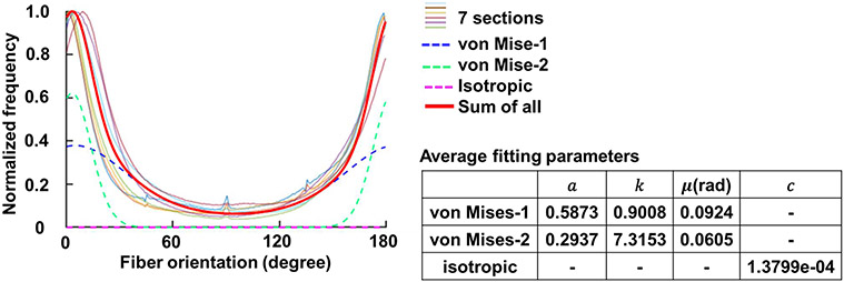
The collagen fibers of the corneoscleral shell play a central role in the eye mechanical behavior. Although it is well-known that these fibers form a complex three-dimensional interwoven structure, biomechanical and microstructural studies often assume that the fibers are aligned in-plane with the tissues. This is convenient as it removes the out-of-plane components and allows focusing on the 2D maps of in-plane fiber organization that are often quite complex. The simplification, however, risks missing potentially important aspects of the tissue architecture and mechanics. In the cornea, for instance, fibers with high in-depth inclination have been shown to be mechanically important. Outside the cornea, the in-depth fiber orientations have not been characterized, preventing a deeper understanding of their potential roles. Our goal was to characterize in-depth collagen fiber organization over the whole corneoscleral shell. Seven sheep whole-globe axial sections from eyes fixed at an IOP of 50 mmHg were imaged using polarized light microscopy to measure collagen fiber orientations and density. In-depth fiber orientation distributions and anisotropy (degree of fiber alignment) accounting for fiber density were quantified over the whole sclera and in 15 regions: central cornea, peripheral cornea, limbus, anterior equator, equator, posterior equator, posterior sclera and peripapillary sclera on both nasal and temporal sides. Orientation distributions were fitted using a combination of a uniform distribution and a sum of π-periodic von Mises distributions, each with three parameters: primary orientation μ, fiber concentration factor k, and weighting factor a. To study the features of fibers that are not in-plane, i.e., fiber inclination, we quantified the percentage of inclined fibers and the range of inclination angles (half width at half maximum of inclination angle distribution). Our measurements showed that the fibers were not uniformly in-plane but exhibited instead a wide range of in-depth orientations, with fibers significantly more aligned in-plane in the anterior parts of the globe. We found that fitting the orientation distributions required between one and three π-periodic von Mises distributions with different primary orientations and fiber concentration factors. Regions of the posterior globe, particularly on the temporal side, had a larger percentage of inclined fibers and a larger range of inclination angles than anterior and equatorial regions. Variations of orientation distributions and anisotropies may imply varying out-of-plane tissue mechanical properties around the eye globe. Out-of-plane fibers could indicate fiber interweaving, not necessarily long, inclined fibers. Effects of small-scale fiber undulations, or crimp, were minimized by using tissues from eyes at high IOPs. These fiber features also play a role in tissue stiffness and stability and are therefore also important experimental information.

摘要

巩膜壳的胶原纤维在眼球的机械行为中起着核心作用。尽管众所周知这些纤维形成了复杂的三维交织结构,但生物力学和微观结构研究通常假设纤维与组织在同一平面内排列。这很方便,因为它消除了非平面分量,并且可以专注于通常非常复杂的平面内纤维组织的 2D 图谱。然而,这种简化可能会忽略组织结构和力学的潜在重要方面。例如,在角膜中,已经证明具有高深度倾斜度的纤维在力学上很重要。在角膜外,尚未对纤维的深度取向进行特征描述,这阻止了对其潜在作用的更深入理解。我们的目标是描述整个巩膜壳中的深度胶原纤维组织。对在 IOP 为 50mmHg 下固定的 7 只绵羊全眼球轴向切片进行偏光显微镜成像,以测量胶原纤维的取向和密度。在整个巩膜和 15 个区域(中央角膜、周边角膜、角膜缘、前赤道、赤道、后赤道、后巩膜和鼻侧和颞侧的视盘周围巩膜)中定量了考虑纤维密度的深度纤维取向分布和各向异性(纤维取向程度)。通过组合使用均匀分布和 π 周期冯·米塞斯(von Mises)分布的总和来拟合取向分布,每个分布具有三个参数:主取向 μ、纤维浓度因子 k 和加权因子 a。为了研究不在同一平面内的纤维特征,即纤维倾斜度,我们定量了倾斜纤维的百分比和倾斜角度范围(倾斜角度分布的半高宽)。我们的测量结果表明,纤维不是均匀地在同一平面内,而是表现出广泛的深度取向,在前部区域的眼球中纤维更平行于平面。我们发现,拟合取向分布需要使用具有不同主取向和纤维浓度因子的一到三个 π 周期冯·米塞斯分布。后半球的区域,特别是颞侧,与前赤道区域相比,倾斜纤维的百分比和倾斜角度范围更大。取向分布和各向异性的变化可能意味着眼球周围的组织具有不同的非平面力学特性。不在同一平面内的纤维可能表示纤维交织,而不一定是长的倾斜纤维。通过使用 IOP 较高的眼睛的组织,可以最小化小尺度纤维波动或卷曲的影响。这些纤维特征在组织刚度和稳定性中也起着重要作用,因此也是重要的实验信息。

https://cdn.ncbi.nlm.nih.gov/pmc/blobs/73f7/10872428/2b178f978678/nihms-1944466-f0011.jpg
https://cdn.ncbi.nlm.nih.gov/pmc/blobs/73f7/10872428/aa6bdc58e135/nihms-1944466-f0001.jpg
https://cdn.ncbi.nlm.nih.gov/pmc/blobs/73f7/10872428/6f4d3160dfbf/nihms-1944466-f0002.jpg
https://cdn.ncbi.nlm.nih.gov/pmc/blobs/73f7/10872428/0ff3d2a8cb26/nihms-1944466-f0003.jpg
https://cdn.ncbi.nlm.nih.gov/pmc/blobs/73f7/10872428/a2ec716f3233/nihms-1944466-f0004.jpg
https://cdn.ncbi.nlm.nih.gov/pmc/blobs/73f7/10872428/f308e1e51d9e/nihms-1944466-f0005.jpg
https://cdn.ncbi.nlm.nih.gov/pmc/blobs/73f7/10872428/eb72bdf1960a/nihms-1944466-f0006.jpg
https://cdn.ncbi.nlm.nih.gov/pmc/blobs/73f7/10872428/431a534e3be8/nihms-1944466-f0007.jpg
https://cdn.ncbi.nlm.nih.gov/pmc/blobs/73f7/10872428/289bac94018e/nihms-1944466-f0008.jpg
https://cdn.ncbi.nlm.nih.gov/pmc/blobs/73f7/10872428/97f159957c19/nihms-1944466-f0009.jpg
https://cdn.ncbi.nlm.nih.gov/pmc/blobs/73f7/10872428/bf580154e39b/nihms-1944466-f0010.jpg
https://cdn.ncbi.nlm.nih.gov/pmc/blobs/73f7/10872428/2b178f978678/nihms-1944466-f0011.jpg

相似文献

1
2D or not 2D? Mapping the in-depth inclination of the collagen fibers of the corneoscleral shell.2D 还是非 2D? 角膜巩膜壳胶原纤维的深度倾斜测绘。
Exp Eye Res. 2023 Dec;237:109701. doi: 10.1016/j.exer.2023.109701. Epub 2023 Oct 26.
2
Who bears the load? IOP-induced collagen fiber recruitment over the corneoscleral shell.谁来承担这个负担?IOP 诱导的角膜巩膜壳上的胶原纤维募集。
Exp Eye Res. 2023 May;230:109446. doi: 10.1016/j.exer.2023.109446. Epub 2023 Mar 18.
3
Crimp around the globe; patterns of collagen crimp across the corneoscleral shell.全球卷曲;角膜巩膜壳上胶原卷曲的模式。
Exp Eye Res. 2018 Jul;172:159-170. doi: 10.1016/j.exer.2018.04.003. Epub 2018 Apr 13.
4
A direct fiber approach to model sclera collagen architecture and biomechanics.一种直接纤维方法来模拟巩膜胶原结构和生物力学。
Exp Eye Res. 2023 Jul;232:109510. doi: 10.1016/j.exer.2023.109510. Epub 2023 May 17.
5
Collagen fiber recruitment: A microstructural basis for the nonlinear response of the posterior pole of the eye to increases in intraocular pressure.胶原纤维募集:眼球后极对眼内压升高的非线性反应的微观结构基础。
Acta Biomater. 2018 May;72:295-305. doi: 10.1016/j.actbio.2018.03.026. Epub 2018 Mar 21.
6
Collagen fiber interweaving is central to sclera stiffness.胶原纤维交织是巩膜硬度的关键。
Acta Biomater. 2020 Sep 1;113:429-437. doi: 10.1016/j.actbio.2020.06.026. Epub 2020 Jun 23.
7
Polarized light microscopy for 3-dimensional mapping of collagen fiber architecture in ocular tissues.用于眼部组织中胶原纤维结构三维映射的偏振光显微镜检查。
J Biophotonics. 2018 Aug;11(8):e201700356. doi: 10.1002/jbio.201700356. Epub 2018 May 6.
8
Collagen Architecture of the Posterior Pole: High-Resolution Wide Field of View Visualization and Analysis Using Polarized Light Microscopy.后极部的胶原结构:使用偏振光显微镜的高分辨率宽视野可视化与分析
Invest Ophthalmol Vis Sci. 2017 Feb 1;58(2):735-744. doi: 10.1167/iovs.16-20772.
9
Spatial Patterns and Age-Related Changes of the Collagen Crimp in the Human Cornea and Sclera.人眼角膜和巩膜胶原卷曲的空间模式和年龄相关性变化。
Invest Ophthalmol Vis Sci. 2018 Jun 1;59(7):2987-2998. doi: 10.1167/iovs.17-23474.
10
Direct measurements of collagen fiber recruitment in the posterior pole of the eye.直接测量眼球后极部的胶原纤维募集。
Acta Biomater. 2024 Jan 1;173:135-147. doi: 10.1016/j.actbio.2023.11.013. Epub 2023 Nov 14.

引用本文的文献

1
Multiscale analysis of equatorial sclera anisotropy: Revealing discrepancies in fiber orientation and mechanical properties.赤道部巩膜各向异性的多尺度分析:揭示纤维取向和力学性能的差异
Sci Adv. 2025 Jul 11;11(28):eadp8631. doi: 10.1126/sciadv.adp8631. Epub 2025 Jul 9.
2
Capturing sclera anisotropy using direct collagen fiber models. Linking microstructure to macroscopic mechanical properties.使用直接胶原纤维模型捕捉巩膜各向异性。将微观结构与宏观力学性能联系起来。
bioRxiv. 2024 Sep 27:2024.09.12.612702. doi: 10.1101/2024.09.12.612702.
3
Comparing continuum and direct fiber models of soft tissues. An ocular biomechanics example reveals that continuum models may artificially disrupt the strains at both the tissue and fiber levels.

本文引用的文献

1
Individual-Specific Modeling of Rat Optic Nerve Head Biomechanics in Glaucoma.青光眼大鼠视神经头生物力学的个体特异性建模。
J Biomech Eng. 2021 Apr 1;143(4). doi: 10.1115/1.4049157.
2
Collagen fiber interweaving is central to sclera stiffness.胶原纤维交织是巩膜硬度的关键。
Acta Biomater. 2020 Sep 1;113:429-437. doi: 10.1016/j.actbio.2020.06.026. Epub 2020 Jun 23.
3
Biomechanical properties of the rat sclera obtained with inverse finite element modeling.利用反有限元建模获得的大鼠巩膜的生物力学特性。
比较软组织的连续体模型和直接纤维模型。一个眼部生物力学实例表明,连续体模型可能会人为地扰乱组织和纤维层面的应变。
bioRxiv. 2024 Sep 10:2024.09.05.610277. doi: 10.1101/2024.09.05.610277.
Biomech Model Mechanobiol. 2020 Dec;19(6):2195-2212. doi: 10.1007/s10237-020-01333-4. Epub 2020 May 2.
4
A Mesh-Free Approach to Incorporate Complex Anisotropic and Heterogeneous Material Properties into Eye-Specific Finite Element Models.一种将复杂各向异性和非均匀材料特性纳入眼部特定有限元模型的无网格方法。
Comput Methods Appl Mech Eng. 2020 Jan 1;358. doi: https://doi.org/10.1016/j.cma.2019.112654. Epub 2019 Oct 1.
5
A Subdomain Method for Mapping the Heterogeneous Mechanical Properties of the Human Posterior Sclera.一种用于绘制人眼后巩膜异质力学特性的子域方法。
Front Bioeng Biotechnol. 2019 May 31;7:129. doi: 10.3389/fbioe.2019.00129. eCollection 2019.
6
Radial and Circumferential Collagen Fibers Are a Feature of the Peripapillary Sclera of Human, Monkey, Pig, Cow, Goat, and Sheep.放射状和环形胶原纤维是人类、猴子、猪、牛、山羊和绵羊的视盘周围巩膜的特征。
Invest Ophthalmol Vis Sci. 2018 Oct 1;59(12):4763-4774. doi: 10.1167/iovs.18-25025.
7
Structured polarized light microscopy for collagen fiber structure and orientation quantification in thick ocular tissues.结构偏振光显微镜用于厚眼部组织中胶原纤维结构和方向的定量分析。
J Biomed Opt. 2018 Oct;23(10):1-10. doi: 10.1117/1.JBO.23.10.106001.
8
Peripapillary sclera architecture revisited: A tangential fiber model and its biomechanical implications.重新探讨视盘周围巩膜结构:一种切向纤维模型及其生物力学意义。
Acta Biomater. 2018 Oct 1;79:113-122. doi: 10.1016/j.actbio.2018.08.020. Epub 2018 Aug 21.
9
Crimp around the globe; patterns of collagen crimp across the corneoscleral shell.全球卷曲;角膜巩膜壳上胶原卷曲的模式。
Exp Eye Res. 2018 Jul;172:159-170. doi: 10.1016/j.exer.2018.04.003. Epub 2018 Apr 13.
10
Polarized light microscopy for 3-dimensional mapping of collagen fiber architecture in ocular tissues.用于眼部组织中胶原纤维结构三维映射的偏振光显微镜检查。
J Biophotonics. 2018 Aug;11(8):e201700356. doi: 10.1002/jbio.201700356. Epub 2018 May 6.