Department of Bioengineering, Swanson School of Engineering, University of Pittsburgh, USA; Department of Ophthalmology, University of Pittsburgh School of Medicine, USA.
Department of Ophthalmology, University of Pittsburgh School of Medicine, USA.
Exp Eye Res. 2018 Jul;172:159-170. doi: 10.1016/j.exer.2018.04.003. Epub 2018 Apr 13.
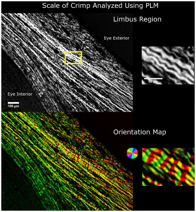
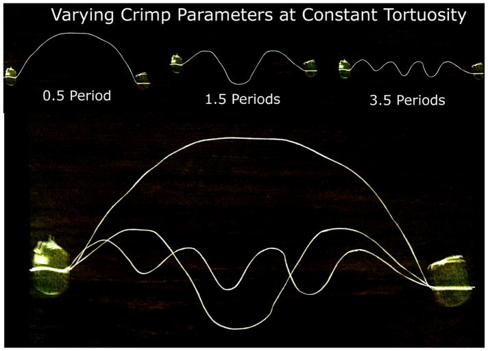
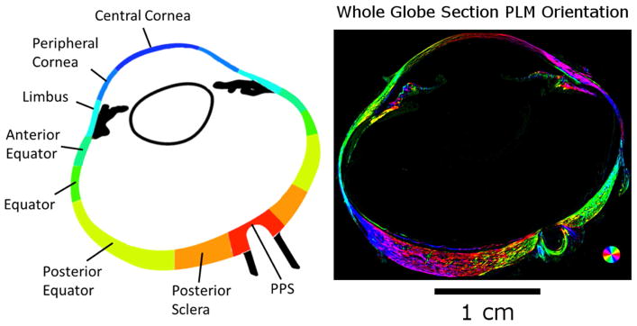
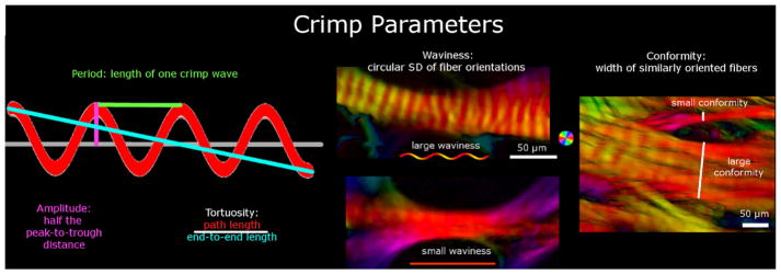
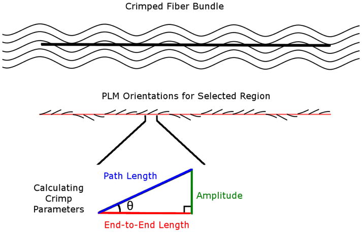
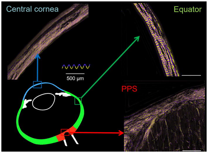
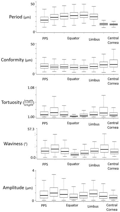
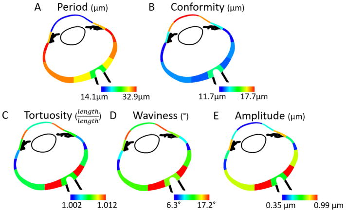
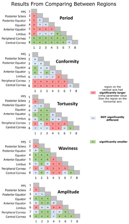
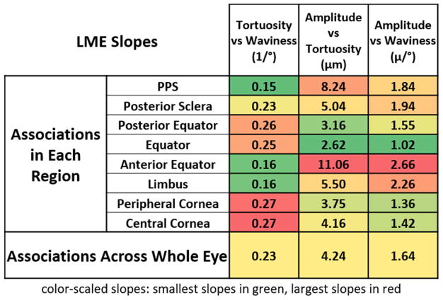
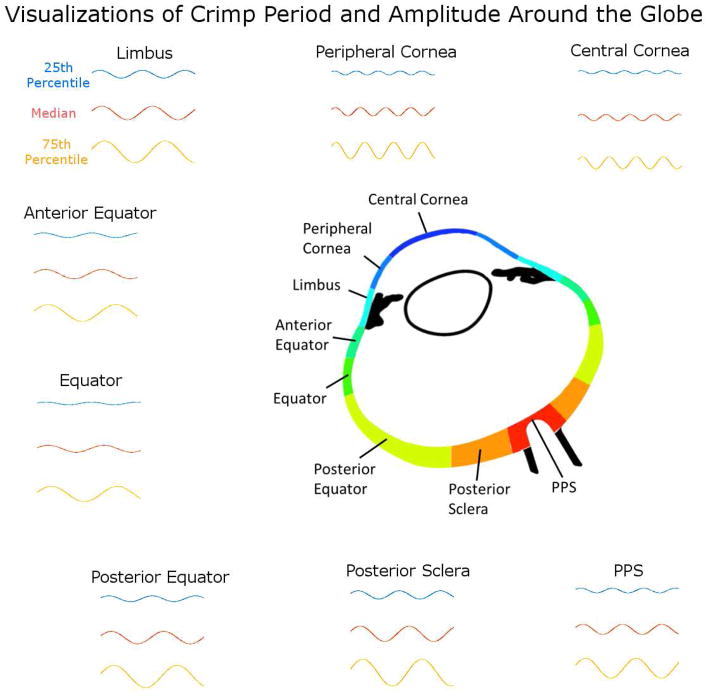
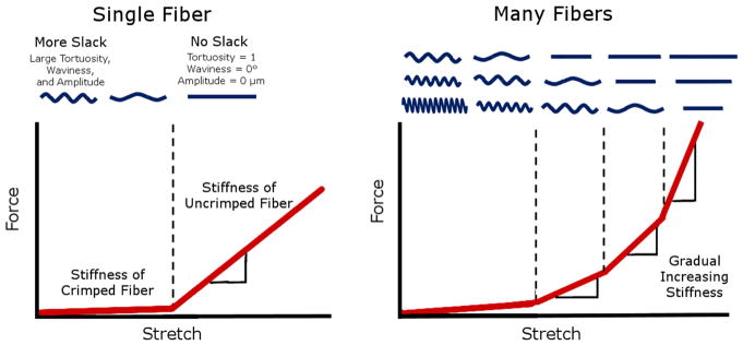
Our goal was to systematically quantify the collagen crimp morphology around the corneoscleral shell, and test the hypothesis that collagen crimp is not uniform over the globe. Axial longitudinal cryosections (30 μm) of three sheep eyes, fixed at 0 mmHg IOP, were imaged using polarized light microscopy to quantify the local collagen in 8 regions: two corneal (central and peripheral) and six scleral (limbus, anterior-equatorial, equatorial, posterior-equatorial, posterior and peripapillary). Collagen crimp period (length of one wave), tortuosity (path length divided by end-to-end length), waviness (SD of orientation), amplitude (half the peak to trough distance), and conformity (width of contiguous similarly oriented bundles) were measured in each region. Measurements were obtained on 8216 collagen fiber bundles. When pooling measurements across the whole eye globe, the median crimp values were: 23.9 μm period, 13.2 μm conformity, 0.63 μm amplitude, 1.006 tortuosity, and 12.7° waviness. However, all parameters varied significantly across the globe. Median bundle periods in the central cornea, limbus, and peripapillary sclera (PPS) were 14.1 μm, 29.5 μm, and 22.9 μm, respectively. Median conformities were 20.8 μm, 14.5 μm, and 15.1 μm, respectively. Median tortuosities were 1.005, 1.007, and 1.007, respectively. Median waviness' were 11.4°, 13.2°, and 13.2°, respectively. Median amplitudes were 0.35 μm, 0.87 μm, and 0.65 μm, respectively. All parameters varied significantly across the globe. All regions differed significantly from one another on at least one parameter. Regions with small periods had large conformities, and bundles with high tortuosity had high waviness and amplitude. Waviness, tortuosity, and amplitude, associated with nonlinear biomechanical behavior, exhibited "double hump" distributions, whereas period and conformity, representing tissue organization, were substantially different between sclera and cornea. Though the biomechanical implications and origin of the patterns observed remain unclear, our findings of well-defined patterns of collagen crimp across the corneoscleral shell, consistent between eyes, support the existence of mechanisms that regulate collagen characteristics at the regional or smaller levels. These results are experimental data necessary for more realistic models of ocular biomechanics and remodeling.
我们的目标是系统地量化角膜巩膜壳周围的胶原卷曲形态,并检验胶原卷曲在整个眼球上不均匀的假设。我们对三只羊眼在眼压为 0mmHg 时固定的轴向纵向冷冻切片(30μm)进行偏振光显微镜成像,以定量测量 8 个区域的局部胶原:两个角膜(中央和周边)和六个巩膜(缘,前赤道,赤道,后赤道,后和视盘周围)。在每个区域测量胶原卷曲周期(一波的长度)、扭曲度(路径长度除以端到端长度)、波纹度(方向的标准差)、振幅(峰谷距离的一半)和一致性(连续相似取向束的宽度)。在 8216 个胶原纤维束上获得测量值。当将整个眼球的测量值汇总时,卷曲值的中位数为:23.9μm 周期、13.2μm 一致性、0.63μm 振幅、1.006 扭曲度和 12.7°波纹度。然而,所有参数在整个眼球上均有显著差异。中央角膜、缘和视盘周围巩膜(PPS)的中位束周期分别为 14.1μm、29.5μm 和 22.9μm。中位一致性分别为 20.8μm、14.5μm 和 15.1μm。中位扭曲度分别为 1.005、1.007 和 1.007。中位波纹度分别为 11.4°、13.2°和 13.2°。中位振幅分别为 0.35μm、0.87μm 和 0.65μm。所有参数在整个眼球上均有显著差异。所有区域在至少一个参数上与其他区域有显著差异。周期较小的区域具有较大的一致性,扭曲度较高的束具有较高的波纹度和振幅。与非线性生物力学行为相关的波纹度、扭曲度和振幅表现出“双峰”分布,而代表组织组织的周期和一致性在巩膜和角膜之间有很大差异。尽管观察到的胶原卷曲模式的生物力学意义和起源仍不清楚,但我们在整个角膜巩膜壳上发现的明确的胶原卷曲模式,以及双眼之间的一致性,支持存在调节区域或更小水平胶原特征的机制。这些结果是眼部生物力学和重塑更现实模型所需的实验数据。