• 文献检索
  • 文档翻译
  • 深度研究
  • 学术资讯
  • Suppr Zotero 插件Zotero 插件
  • 邀请有礼
  • 套餐&价格
  • 历史记录
应用&插件
Suppr Zotero 插件Zotero 插件浏览器插件Mac 客户端Windows 客户端微信小程序
定价
高级版会员购买积分包购买API积分包
服务
文献检索文档翻译深度研究API 文档MCP 服务
关于我们
关于 Suppr公司介绍联系我们用户协议隐私条款
关注我们

Suppr 超能文献

核心技术专利:CN118964589B侵权必究
粤ICP备2023148730 号-1Suppr @ 2026

文献检索

告别复杂PubMed语法,用中文像聊天一样搜索,搜遍4000万医学文献。AI智能推荐,让科研检索更轻松。

立即免费搜索

文件翻译

保留排版,准确专业,支持PDF/Word/PPT等文件格式,支持 12+语言互译。

免费翻译文档

深度研究

AI帮你快速写综述,25分钟生成高质量综述,智能提取关键信息,辅助科研写作。

立即免费体验

数字乳腺断层合成的乳腺密度分析。

Breast density analysis of digital breast tomosynthesis.

机构信息

Cancer Epidemiology Department, Moffitt Cancer Center and Research Institute, 12902 Bruce B. Downs Blvd, Tampa, FL, 33612, USA.

Diagnostic Imaging and Interventional Radiology, Moffitt Cancer Center and Research Institute, 12902 Bruce B. Downs Blvd, Tampa, FL, 33612, USA.

出版信息

Sci Rep. 2023 Oct 31;13(1):18760. doi: 10.1038/s41598-023-45402-x.

DOI:10.1038/s41598-023-45402-x
PMID:37907569
原文链接:https://pmc.ncbi.nlm.nih.gov/articles/PMC10618274/
Abstract

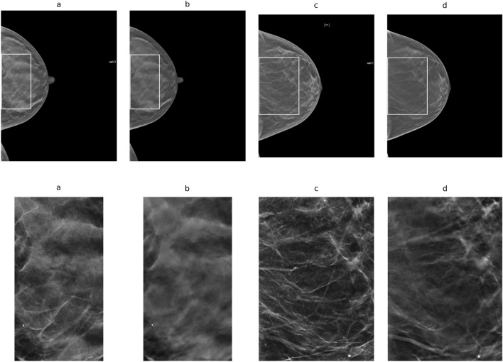
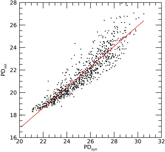
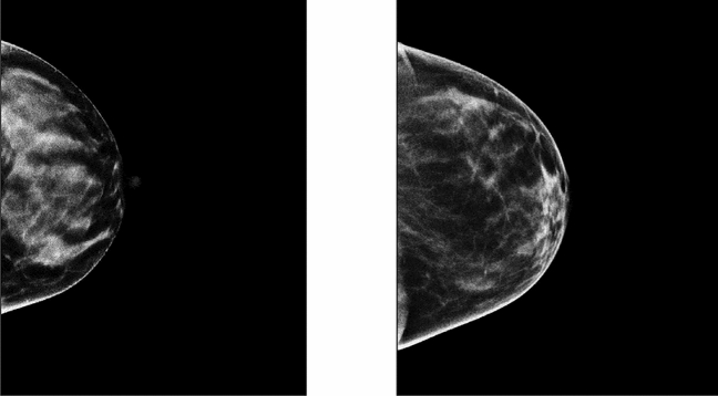
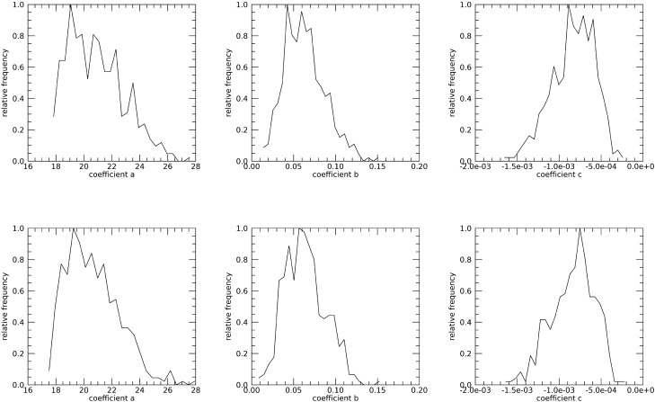
Mammography shifted to digital breast tomosynthesis (DBT) in the US. An automated percentage of breast density (PD) technique designed for two-dimensional (2D) applications was evaluated with DBT using several breast cancer risk prediction measures: normalized-volumetric; dense volume; applied to the volume slices and averaged (slice-mean); and applied to synthetic 2D images. Volumetric measures were derived theoretically. PD was modeled as a function of compressed breast thickness (CBT). The mean and standard deviation of the pixel values were investigated. A matched case-control (CC) study (n = 426 pairs) was evaluated. Odd ratios (ORs) were estimated with 95% confidence intervals. ORs were significant for PD: identical for volumetric and slice-mean measures [OR = 1.43 (1.18, 1.72)] and [OR = 1.44 (1.18, 1.75)] for synthetic images. A 2nd degree polynomial (concave-down) was used to model PD as a function of CBT: location of the maximum PD value was similar across CCs, occurring at 0.41 × CBT, and PD was significant [OR = 1.47 (1.21, 1.78)]. The means from the volume and synthetic images were also significant [ORs ~ 1.31 (1.09, 1.57)]. An alternative standardized 2D synthetic image was constructed, where each pixel value represents the percentage of breast density above its location. Several measures were significant and an alternative method for constructing a standardized 2D synthetic image was produced.

摘要

在美国,乳腺 X 光摄影已转向数字乳腺断层合成技术(DBT)。评估了一种针对二维(2D)应用设计的自动乳腺密度(PD)百分比技术,该技术使用了几种乳腺癌风险预测指标:归一化体积;致密体积;应用于体积切片并取平均值(切片平均值);以及应用于合成 2D 图像。体积测量是从理论上推导出来的。PD 被建模为压缩乳腺厚度(CBT)的函数。研究了像素值的平均值和标准差。评估了一项匹配病例对照(CC)研究(n=426 对)。估计了 95%置信区间的比值比(OR)。PD 的 OR 具有统计学意义:体积和切片平均值测量的 OR 相同[OR=1.43(1.18,1.72)]和[OR=1.44(1.18,1.75)]对于合成图像。使用二次多项式(凹向下)将 PD 建模为 CBT 的函数:CC 之间最大 PD 值的位置相似,出现在 0.41×CBT 处,PD 有统计学意义[OR=1.47(1.21,1.78)]。体积和合成图像的平均值也具有统计学意义[ORs~1.31(1.09,1.57)]。还构建了替代的标准化 2D 合成图像,其中每个像素值代表其位置以上的乳腺密度百分比。有几个指标具有统计学意义,并生成了构建标准化 2D 合成图像的替代方法。

https://cdn.ncbi.nlm.nih.gov/pmc/blobs/17d9/10618274/d4c5590503da/41598_2023_45402_Fig7_HTML.jpg
https://cdn.ncbi.nlm.nih.gov/pmc/blobs/17d9/10618274/5e7757fc733d/41598_2023_45402_Fig1_HTML.jpg
https://cdn.ncbi.nlm.nih.gov/pmc/blobs/17d9/10618274/8801b35dd1b0/41598_2023_45402_Fig2_HTML.jpg
https://cdn.ncbi.nlm.nih.gov/pmc/blobs/17d9/10618274/bbf19273c4a4/41598_2023_45402_Fig3_HTML.jpg
https://cdn.ncbi.nlm.nih.gov/pmc/blobs/17d9/10618274/6a0b8348161b/41598_2023_45402_Fig4_HTML.jpg
https://cdn.ncbi.nlm.nih.gov/pmc/blobs/17d9/10618274/79f60208b639/41598_2023_45402_Fig5_HTML.jpg
https://cdn.ncbi.nlm.nih.gov/pmc/blobs/17d9/10618274/3ef8b42d1b90/41598_2023_45402_Fig6_HTML.jpg
https://cdn.ncbi.nlm.nih.gov/pmc/blobs/17d9/10618274/d4c5590503da/41598_2023_45402_Fig7_HTML.jpg

相似文献

1
Breast density analysis of digital breast tomosynthesis.数字乳腺断层合成的乳腺密度分析。
Sci Rep. 2023 Oct 31;13(1):18760. doi: 10.1038/s41598-023-45402-x.
2
Breast Density Analysis Using Digital Breast Tomosynthesis.使用数字乳腺断层合成技术进行乳腺密度分析。
bioRxiv. 2023 Feb 16:2023.02.10.527911. doi: 10.1101/2023.02.10.527911.
3
Diagnostic accuracy of contrast-enhanced digital mammography in breast cancer detection in comparison to tomosynthesis, synthetic 2D mammography and tomosynthesis combined with ultrasound in women with dense breast.与断层合成、合成二维乳腺摄影以及断层合成联合超声相比,对比增强数字乳腺摄影在致密型乳腺女性乳腺癌检测中的诊断准确性。
Br J Radiol. 2021 Feb 1;94(1118):20201046. doi: 10.1259/bjr.20201046. Epub 2020 Dec 2.
4
Comparing recall rates following implementation of digital breast tomosynthesis to synthetic 2D images and digital mammography on women with breast-conserving surgery.比较保乳手术后数字乳腺断层合成影像和数字乳腺钼靶对召回率的影响。
Eur Radiol. 2020 Nov;30(11):6072-6079. doi: 10.1007/s00330-020-06992-6. Epub 2020 Jun 11.
5
Radiation dose with digital breast tomosynthesis compared to digital mammography: per-view analysis.数字乳腺断层合成与数字乳腺钼靶摄影的放射剂量比较:单视图分析。
Eur Radiol. 2018 Feb;28(2):573-581. doi: 10.1007/s00330-017-5024-4. Epub 2017 Aug 17.
6
Automated Breast Density Computation in Digital Mammography and Digital Breast Tomosynthesis: Influence on Mean Glandular Dose and BIRADS Density Categorization.数字化乳腺摄影和数字乳腺断层合成中的自动乳腺密度计算:对平均腺体剂量和乳腺影像报告和数据系统(BIRADS)密度分类的影响
Acad Radiol. 2017 Jul;24(7):802-810. doi: 10.1016/j.acra.2017.01.011. Epub 2017 Feb 14.
7
8
Fully Automated Volumetric Breast Density Estimation from Digital Breast Tomosynthesis.从数字乳腺断层合成术中全自动体积乳腺密度估计
Radiology. 2021 Dec;301(3):561-568. doi: 10.1148/radiol.2021210190. Epub 2021 Sep 14.
9
Estimation of percentage breast tissue density: comparison between digital mammography (2D full field digital mammography) and digital breast tomosynthesis according to different BI-RADS categories.乳腺组织密度百分比的估计:数字乳腺钼靶摄影(二维全视野数字化乳腺钼靶摄影)与数字乳腺断层合成技术在不同 BI-RADS 分类中的比较。
Br J Radiol. 2013 Nov;86(1031):20130255. doi: 10.1259/bjr.20130255. Epub 2013 Sep 12.
10
Evaluation of LIBRA Software for Fully Automated Mammographic Density Assessment in Breast Cancer Risk Prediction.评估 LIBRA 软件在乳腺癌风险预测中全自动乳腺密度评估的应用。
Radiology. 2020 Jul;296(1):24-31. doi: 10.1148/radiol.2020192509. Epub 2020 May 12.

引用本文的文献

1
Cross-field strength and multi-vendor reliability of MagDensity for MRI-based quantitative breast density analysis.用于基于MRI的乳腺密度定量分析的磁密度的跨场强度和多供应商可靠性。
PLoS One. 2025 Jun 24;20(6):e0316076. doi: 10.1371/journal.pone.0316076. eCollection 2025.
2
Fourier analysis of signal dependent noise images.信号相关噪声图像的傅里叶分析
Sci Rep. 2024 Dec 28;14(1):30686. doi: 10.1038/s41598-024-78299-1.
3
Breast Cancer Screening and Prophylactic Mastectomy for High-Risk Women in Romania.罗马尼亚高危女性的乳腺癌筛查与预防性乳房切除术

本文引用的文献

1
Breast Cancer Risk Assessment: A Review on Mammography-Based Approaches.乳腺癌风险评估:基于乳腺X线摄影方法的综述
J Imaging. 2021 Jun 12;7(6):98. doi: 10.3390/jimaging7060098.
2
Mammographic breast density and the risk of breast cancer: A systematic review and meta-analysis.乳腺钼靶密度与乳腺癌风险:系统评价和荟萃分析。
Breast. 2022 Dec;66:62-68. doi: 10.1016/j.breast.2022.09.007. Epub 2022 Sep 26.
3
A risk model for digital breast tomosynthesis to predict breast cancer and guide clinical care.一种用于数字乳腺断层合成的风险模型,以预测乳腺癌并指导临床护理。
Medicina (Kaunas). 2024 Mar 30;60(4):570. doi: 10.3390/medicina60040570.
Sci Transl Med. 2022 May 11;14(644):eabn3971. doi: 10.1126/scitranslmed.abn3971.
4
Fabrication of 3D printed patient-derived anthropomorphic breast phantoms for mammography and digital breast tomosynthesis: Imaging assessment with clinical X-ray spectra.使用临床 X 射线光谱对用于乳腺 X 线摄影和数字乳腺断层合成的 3D 打印患者衍生拟人化乳房体模的制作进行成像评估。
Phys Med. 2022 Jun;98:88-97. doi: 10.1016/j.ejmp.2022.04.006. Epub 2022 May 5.
5
Artificial intelligence in mammographic phenotyping of breast cancer risk: a narrative review.人工智能在乳腺癌风险的乳腺摄影表型中的应用:叙述性综述。
Breast Cancer Res. 2022 Feb 20;24(1):14. doi: 10.1186/s13058-022-01509-z.
6
ACR Appropriateness Criteria® Supplemental Breast Cancer Screening Based on Breast Density.ACR 适宜性标准®基于乳腺密度的补充乳腺癌筛查。
J Am Coll Radiol. 2021 Nov;18(11S):S456-S473. doi: 10.1016/j.jacr.2021.09.002.
7
Fully Automated Volumetric Breast Density Estimation from Digital Breast Tomosynthesis.从数字乳腺断层合成术中全自动体积乳腺密度估计
Radiology. 2021 Dec;301(3):561-568. doi: 10.1148/radiol.2021210190. Epub 2021 Sep 14.
8
Mammographic Variation Measures, Breast Density, and Breast Cancer Risk.乳腺 X 线摄影变异指标、乳腺密度与乳腺癌风险
AJR Am J Roentgenol. 2021 Aug;217(2):326-335. doi: 10.2214/AJR.20.22794. Epub 2021 Jun 23.
9
Automated percent mammographic density, mammographic texture variation, and risk of breast cancer: a nested case-control study.乳腺X线摄影密度自动化百分比、乳腺X线摄影纹理变化与乳腺癌风险:一项巢式病例对照研究
NPJ Breast Cancer. 2021 May 31;7(1):68. doi: 10.1038/s41523-021-00272-2.
10
Spatial Correlation and Breast Cancer Risk.空间相关性与乳腺癌风险
Biomed Phys Eng Express. 2019 Jul;5(4). doi: 10.1088/2057-1976/ab1dad. Epub 2019 May 22.