Tison Geoffrey H, Abreau Sean, Barrios Joshua, Lim Lisa J, Yang Michelle, Crudo Valentina, Shah Dipan J, Nguyen Thuy, Hu Gene, Dixit Shalini, Nah Gregory, Arya Farzin, Bibby Dwight, Lee Yoojin, Delling Francesca N
Cardiovascular Division, Department of Medicine, University of California-San Francisco, San Francisco, California, USA.
Bakar Computational Health Sciences Institute, University of California, San Francisco, California, USA.
JACC Adv. 2023 Aug;2(6). doi: 10.1016/j.jacadv.2023.100446. Epub 2023 Aug 5.
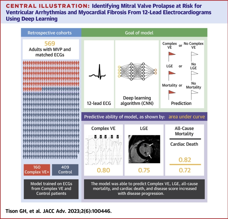
Mitral valve prolapse (MVP) is a common valvulopathy, with a subset developing sudden cardiac death or cardiac arrest. Complex ventricular ectopy (ComVE) is a marker of arrhythmic risk associated with myocardial fibrosis and increased mortality in MVP.
The authors sought to evaluate whether electrocardiogram (ECG)-based machine learning can identify MVP at risk for ComVE, death and/or myocardial fibrosis on cardiac magnetic resonance (CMR) imaging.
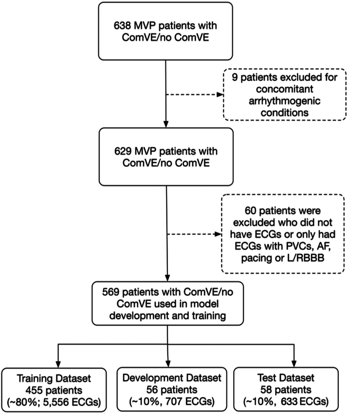
A deep convolutional neural network (CNN) was trained to detect ComVE using 6,916 12-lead ECGs from 569 MVP patients from the University of California-San Francisco between 2012 and 2020. A separate CNN was trained to detect late gadolinium enhancement (LGE) using 1,369 ECGs from 87 MVP patients with contrast CMR.
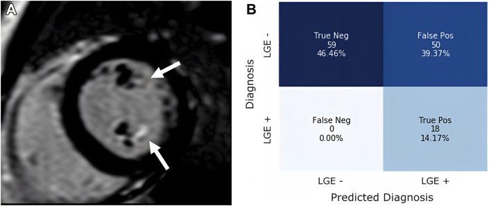
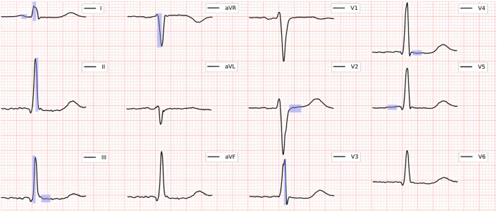
The prevalence of ComVE was 28% (160/569). The area under the receiver operating characteristic curve (AUC) of the CNN to detect ComVE was 0.80 (95% CI: 0.77-0.83) and remained high after excluding patients with moderate-severe mitral regurgitation [0.80 (95% CI: 0.77-0.83)] or bileaflet MVP [0.81 (95% CI: 0.76-0.85)]. AUC to detect all-cause mortality was 0.82 (95% CI: 0.77-0.87). ECG segments relevant to ComVE prediction were related to ventricular depolarization/repolarization (early-mid ST-segment and QRS from V, V, and III). LGE in the papillary muscles or basal inferolateral wall was present in 24% patients with available CMR; AUC for detection of LGE was 0.75 (95% CI: 0.68-0.82).
CNN-analyzed 12-lead ECGs can detect MVP at risk for ventricular arrhythmias, death and/or fibrosis and can identify novel ECG correlates of arrhythmic risk. ECG-based CNNs may help select those MVP patients requiring closer follow-up and/or a CMR.
二尖瓣脱垂(MVP)是一种常见的瓣膜病,其中一部分患者会发生心源性猝死或心脏骤停。复杂室性早搏(ComVE)是与心肌纤维化相关的心律失常风险标志物,且在MVP患者中死亡率增加。
作者试图评估基于心电图(ECG)的机器学习能否识别有ComVE、死亡和/或心脏磁共振成像(CMR)显示心肌纤维化风险的MVP患者。
使用来自加利福尼亚大学旧金山分校2012年至2020年期间569例MVP患者的6916份12导联心电图,训练一个深度卷积神经网络(CNN)来检测ComVE。使用来自87例进行对比CMR检查的MVP患者的1369份心电图,训练另一个单独的CNN来检测延迟钆增强(LGE)。
ComVE的患病率为28%(160/569)。用于检测ComVE的CNN的受试者工作特征曲线下面积(AUC)为0.80(95%CI:0.77-0.83),在排除中重度二尖瓣反流患者[0.80(95%CI:0.77-0.83)]或双叶MVP患者[0.81(95%CI:0.76-0.85)]后仍保持较高水平。检测全因死亡率的AUC为0.82(95%CI:0.77-0.87)。与ComVE预测相关的ECG节段与心室去极化/复极化有关(V、V和III导联的ST段早期至中期及QRS波)。在有CMR检查结果的患者中,24%的患者乳头肌或基底下侧壁存在LGE;检测LGE的AUC为0.75(95%CI:0.68-0.82)。
经CNN分析的12导联心电图可检测有室性心律失常、死亡和/或纤维化风险的MVP患者,并可识别心律失常风险的新ECG相关因素。基于ECG的CNN可能有助于选择那些需要密切随访和/或进行CMR检查的MVP患者。