Qian Ming, Chen Xi, Zhang Long-Yao, Wang Zhi-Feng, Zhang Yi, Wang Xue-Jian
Department of Neurosurgery, Affiliated Hospital 2 of Nantong University, Nantong 226000, Jiangsu Province, China.
Department of Nursing, Affiliated Hospital 2 of Nantong University, Nantong 226001, Jiangsu Province, China.
World J Clin Cases. 2023 Oct 16;11(29):7053-7060. doi: 10.12998/wjcc.v11.i29.7053.
At present, neuroendoscopy technology has made rapid development, and great progress has been made in the operation of lesions in the saddle area of the skull base. However, the complications of cerebrospinal fluid and intracranial infection after the operation are still important and life-threatening complications, which may lead to poor prognosis.
To investigate the method of in situ bone flap combined with nasal septum mucosal flap for reconstruction of enlarged skull base defect by endonasal sphenoidal approach and to discuss its application effect.
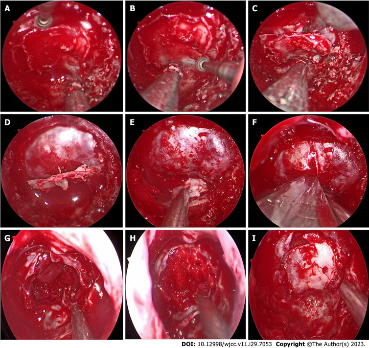
Clinical data of 24 patients undergoing transnasal sphenoidal endoscopic approach in the Department of Neurosurgery, Affiliated 2 Hospital of Nantong University from January 2019 to December 2022 were retrospectively analyzed. All patients underwent multi-layer reconstruction of skull base using in situ bone flap combined with nasal septum mucosa flap. The incidence of intraoperative and postoperative cerebrospinal fluid leakage and intracranial infection were analyzed, and the application effect and technical key points of in situ bone flap combined with nasal septum mucosa flap for skull base bone reconstruction were analyzed.
There were 5 cases of high flow cerebrospinal fluid (CSF) leakage and 7 cases of low flow CSF leakage. Postoperative cerebrospinal fluid leakage occurred in 2 patients (8.3%) and intracranial infection in 2 patients (8.3%), which were cured after strict bed rest, continuous drainage of lumbar cistern combined with antibiotic treatment, and no secondary surgical repair was required. The patients were followed up for 8 to 36 months after the operation, and no delayed cerebrospinal fluid leakage or intracranial infection occurred during the follow-up. Computed tomography reconstruction of skull base showed satisfactory reconstruction after surgery.
The use of in situ bone flap combined with vascular pedicled mucous flap to reconstruction of skull base defect after endonasal sphenoidal approach under neuroendoscopy has a lower incidence of cerebrospinal fluid leakage and lower complications, which has certain advantages and is worthy of clinical promotion.
目前,神经内镜技术发展迅速,在颅底鞍区病变的手术治疗方面取得了很大进展。然而,术后脑脊液及颅内感染等并发症仍是重要的、危及生命的并发症,可能导致预后不良。
探讨经鼻蝶入路原位骨瓣联合鼻中隔黏膜瓣修复扩大的颅底缺损的方法,并探讨其应用效果。
回顾性分析2019年1月至2022年12月南通大学第二附属医院神经外科收治的24例行经鼻蝶内镜手术患者的临床资料。所有患者均采用原位骨瓣联合鼻中隔黏膜瓣进行颅底多层重建。分析术中及术后脑脊液漏和颅内感染的发生率,分析原位骨瓣联合鼻中隔黏膜瓣用于颅底骨重建的应用效果及技术要点。
高流量脑脊液漏5例,低流量脑脊液漏7例。术后2例(8.3%)发生脑脊液漏,2例(8.3%)发生颅内感染,经严格卧床休息、腰大池持续引流联合抗生素治疗后治愈,无需二次手术修复。术后随访8~36个月,随访期间无迟发性脑脊液漏及颅内感染发生。颅底CT重建显示术后重建效果满意。
神经内镜下经鼻蝶入路术后采用原位骨瓣联合带血管蒂黏膜瓣修复颅底缺损,脑脊液漏发生率低,并发症少,具有一定优势,值得临床推广。