Siemens Medical Solutions, Knoxville, Tennessee, USA.
Department of Radiology, Perelman School of Medicine, University of Pennsylvania, Philadelphia, Pennsylvania, USA.
Med Phys. 2024 Jan;51(1):54-69. doi: 10.1002/mp.16826. Epub 2023 Nov 13.
Scatter correction (SC) is essential in PET for accurate quantitative imaging. The state-of-the-art SC method is single-scatter simulation (SSS). Although this method is usually robust and accurate, it can fail in some situations, for example when there is motion between the CT and PET scans in PET/CT. Therefore, it is of interest to consider other SC methods.
In this work, an energy-based scatter estimation (EBS) method is described in detail, tested in phantoms and patients, and compared to SSS.
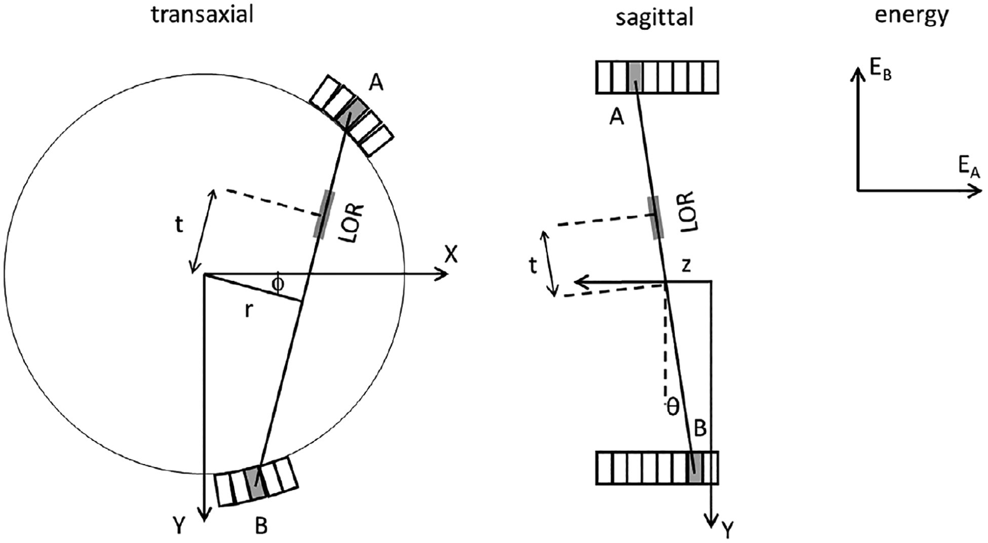
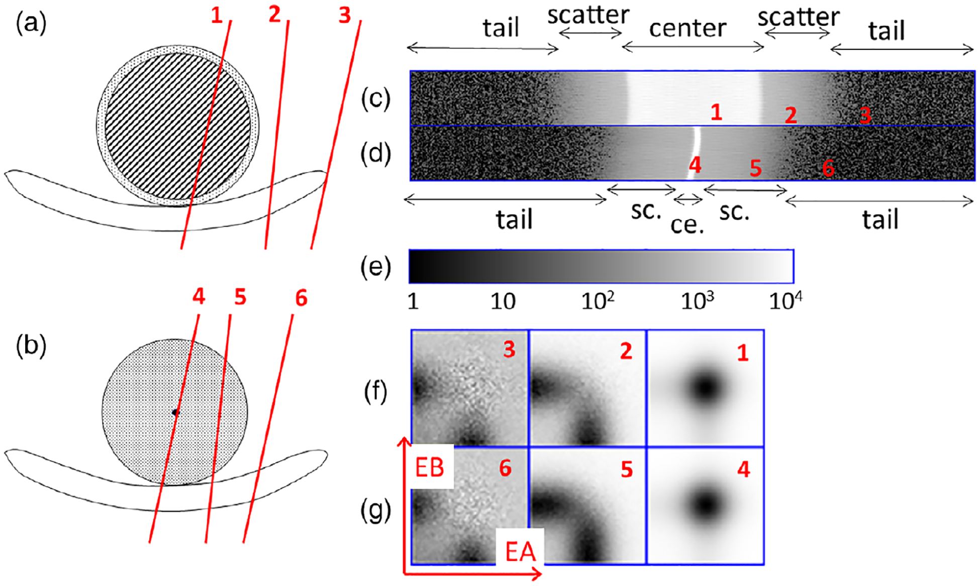
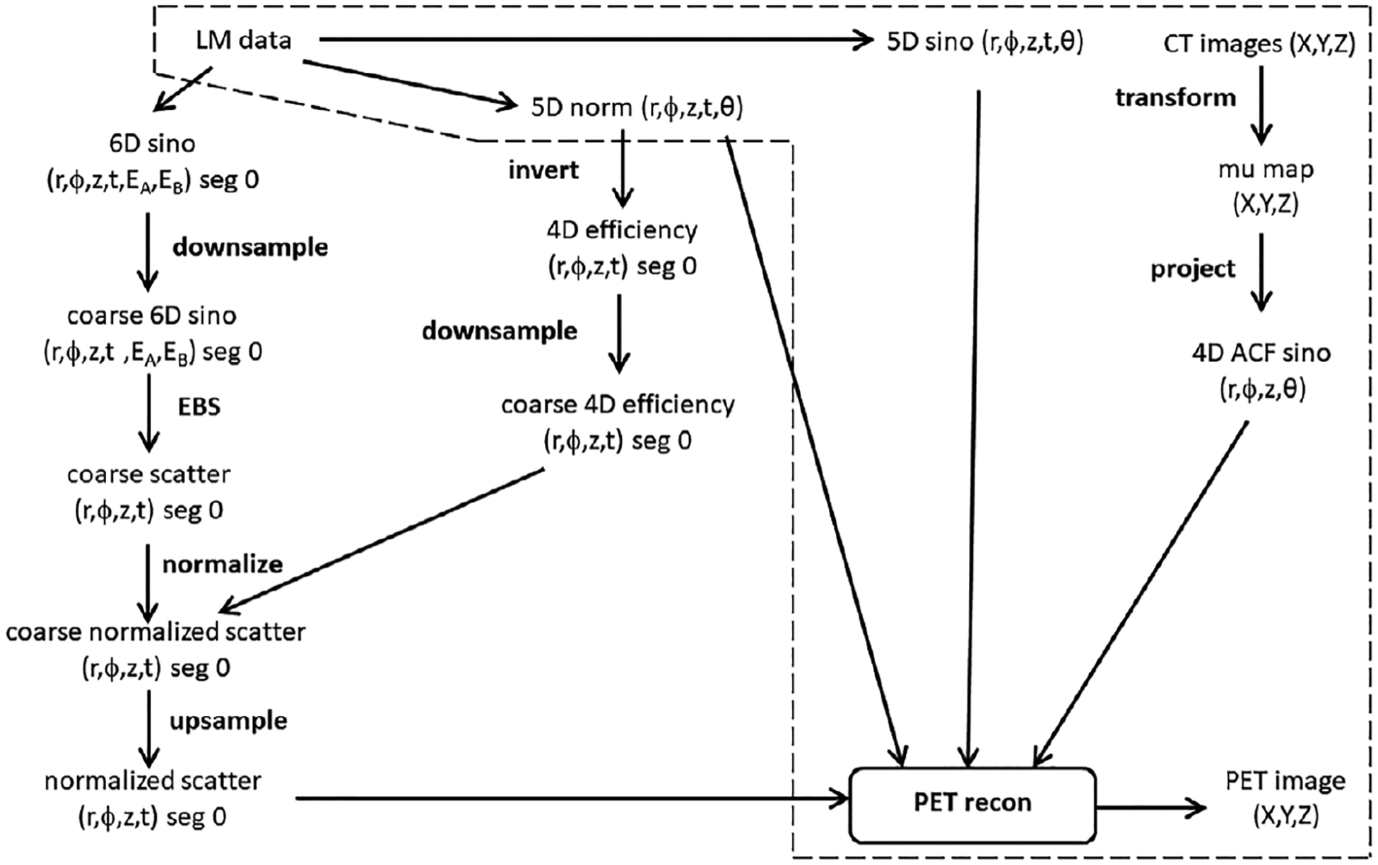
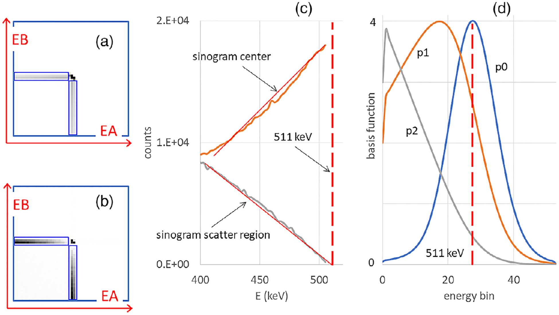
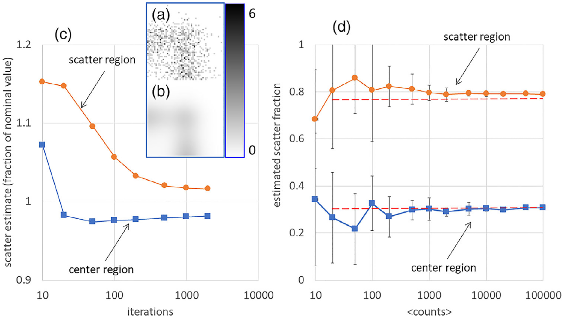
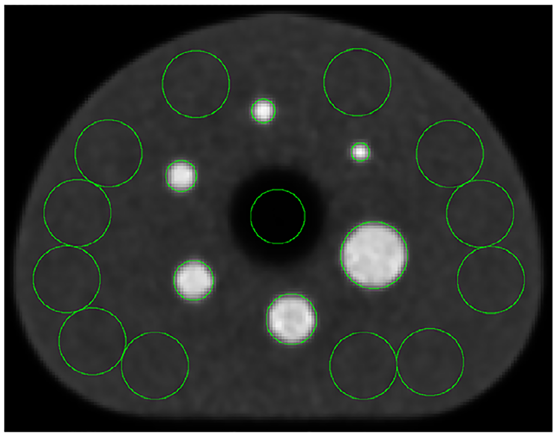
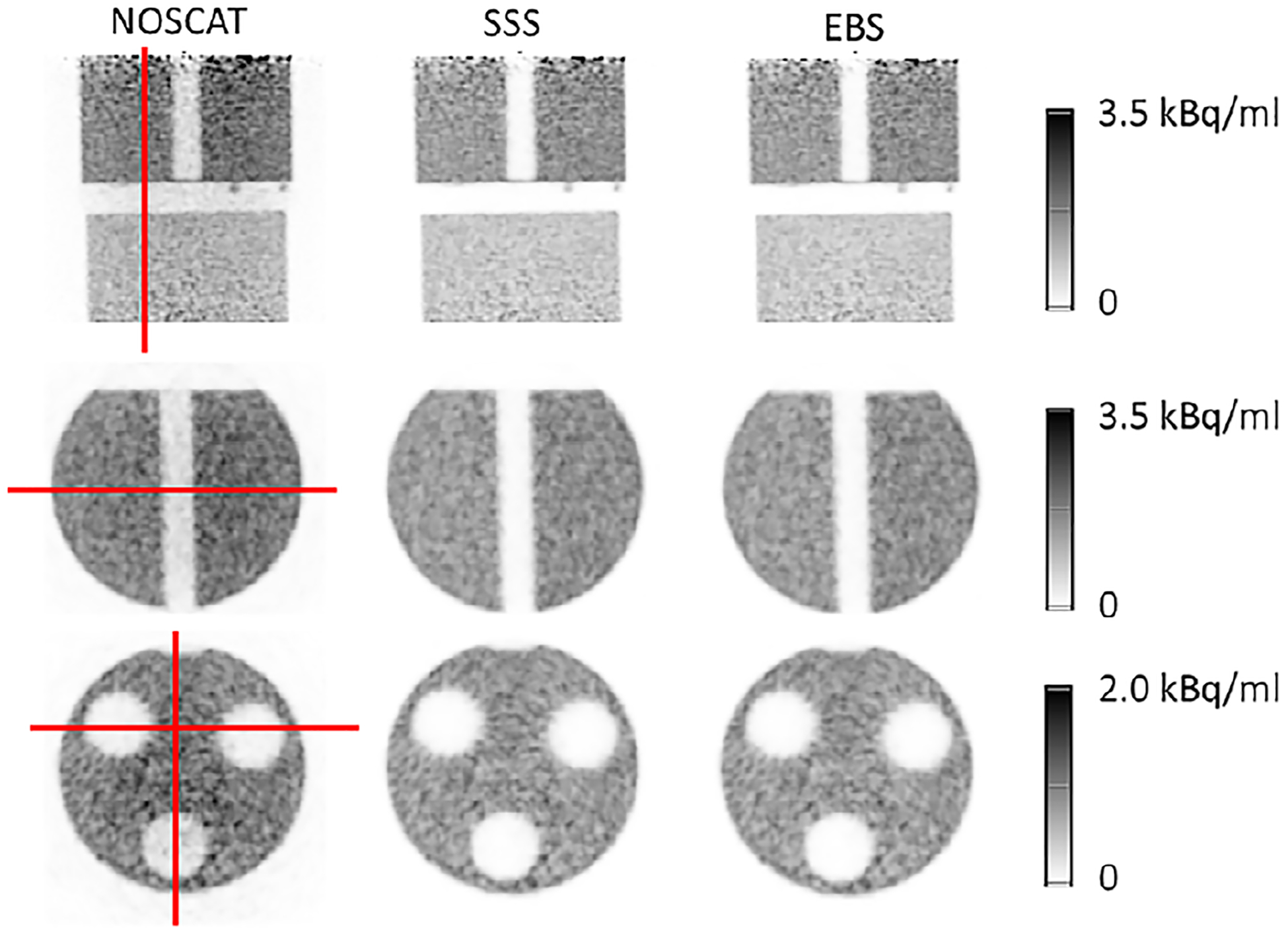
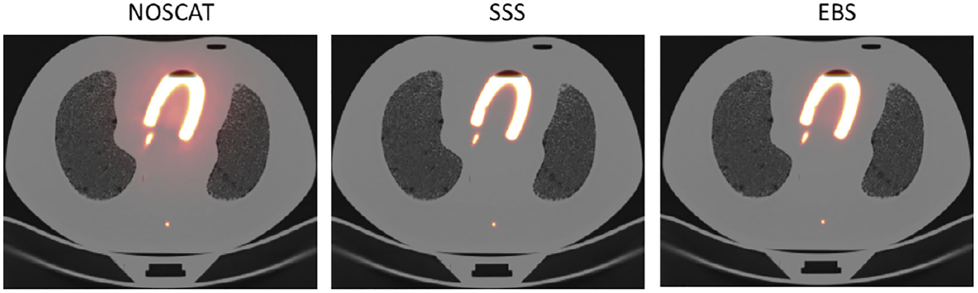
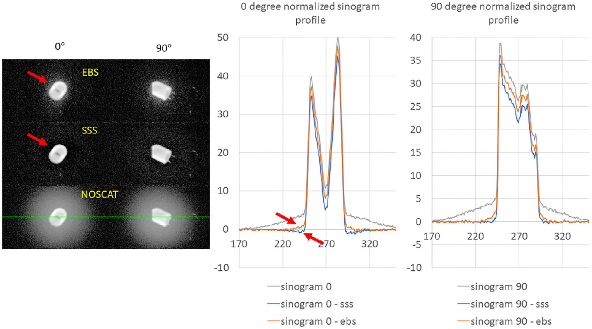
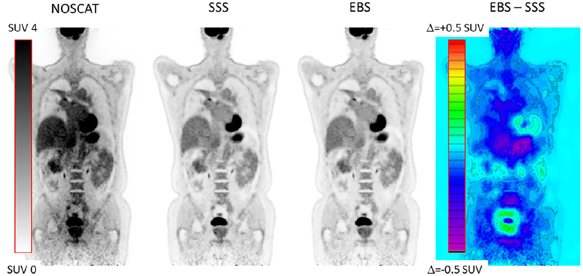
This version of EBS was developed for list-mode data from Biograph Vision-600 PET/CT scanner. EBS is based on digitized 2D energy histograms in each bin of a coarsely sampled PET sinogram, either with or without time of flight (TOF). The histograms are modeled as a noisy realization of a linear combination of nine basis functions whose parameters were derived from a measurement of the 511-keV photopeak spectrum as well as Monte-Carlo simulations of the scattering process. EBS uses an iterative expectation maximization approach to determine the coefficients in the linear combination, and from this estimates the scatter. The investigation was restricted to F-based PET data in which the acquired number of counts was similar to the levels seen in oncological whole-body PET/CT scans. To evaluate the performance, phantom scans were used that involved the NEMA NU2-2018 protocol, a slab phantom, an NU 2-1994 phantom, a cardiac phantom in an anthropomorphic chest phantom, and a uniformly-filled torso phantom with a bladder phantom slightly outside the axial field of view. Contrast recovery (CR) and other parameters were evaluated in images reconstructed with SSS and EBS. Furthermore, FDG PET scans of seven lung cancer patients were used in the evaluation. Standardized uptake values (SUV) based on SSS and EBS were compared in 27 lesions.
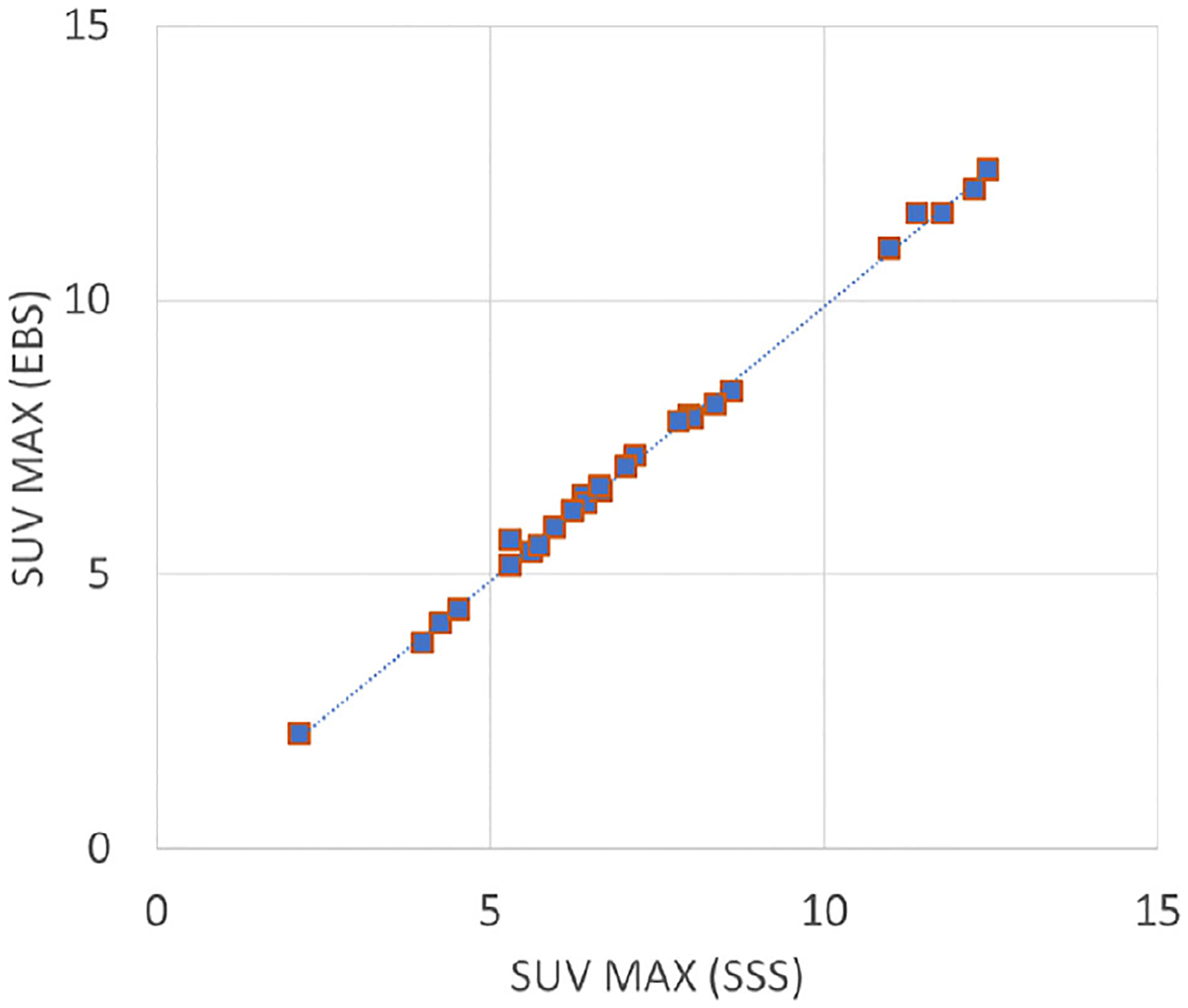
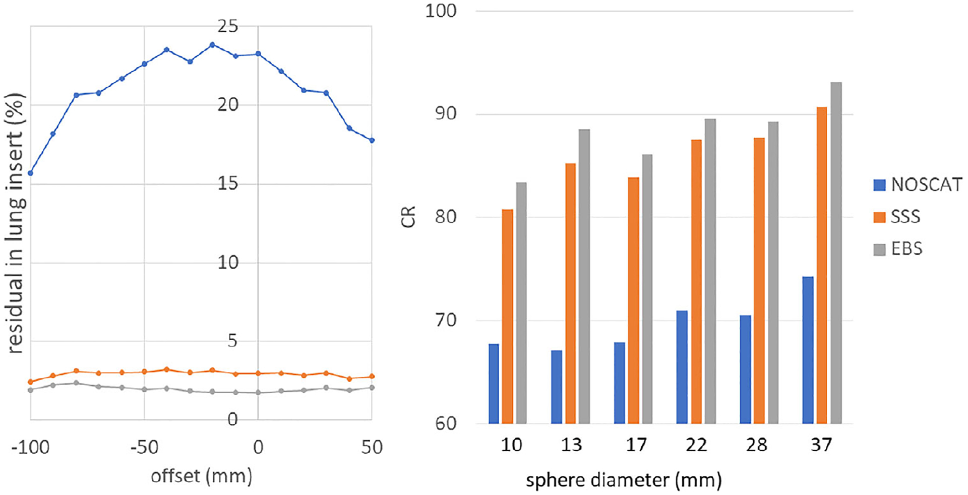
EBS and SSS images were visually similar in all cases except the torso + bladder phantom, where the EBS was much closer to the expected uniform image. The NU2-2018 analysis indicated a 2% scatter residual in EBS images compared to 3% with SSS, and 10% higher background variability, which is a surrogate for image noise. The cardiac phantom scan showed that CR was 98.2% with EBS and 99.6% with SSS, and that the SSS sinogram had values greater than the net-true emission sinogram, indicating a slight overcorrection in the case of SSS. In the lesion SUV comparison in patient scans, EBS correlated strongly (R = 0.9973) with SSS, and SUV based on EBS were systematically 0.1 SUV lower. In the case of the torso + bladder phantom portion, the SSS image of the torso + bladder phantom was 299% times hotter than expected in one area, due to scatter estimation error, compared to 16% colder with EBS.
In evaluating clinically relevant parameters such as SUV in focal lesions, EBS and SSS give almost the same results. In phantoms, some scatter figures of merit were slightly improved by use of EBS, though an image variability figure of merit was slightly degraded. In typical oncological whole-body PET/CT, EBS may be a suitable replacement for SSS, especially when SSS fails due to technical problems during the scan.
在 PET 中,散射校正(SC)对于准确的定量成像至关重要。最先进的 SC 方法是单散射模拟(SSS)。尽管这种方法通常是稳健和准确的,但在某些情况下可能会失败,例如在 PET/CT 中 CT 和 PET 扫描之间存在运动时。因此,考虑其他 SC 方法是很有意义的。
在这项工作中,详细描述了一种基于能量的散射估计(EBS)方法,并在体模和患者中进行了测试,并与 SSS 进行了比较。
这个版本的 EBS 是为 Biograph Vision-600 PET/CT 扫描仪的列表模式数据开发的。EBS 基于在粗采样的 PET 正弦图的每个 bin 中数字化的 2D 能量直方图,无论是否具有飞行时间(TOF)。直方图被建模为一个线性组合的噪声实现,其参数是从 511keV 光峰谱的测量以及散射过程的蒙特卡罗模拟中得出的。EBS 使用迭代期望最大化方法来确定线性组合中的系数,并从这个估计散射。研究仅限于 F 型 PET 数据,其中采集的计数数与肿瘤全身 PET/CT 扫描中看到的计数数相似。为了评估性能,使用了体模扫描,包括 NEMA NU2-2018 协议、平板体模、NU 2-1994 体模、在人体胸部体模中的心脏体模以及充满均匀物质的躯干体模,其中膀胱体模稍微超出轴向视野。在使用 SSS 和 EBS 重建的图像中评估了对比度恢复(CR)和其他参数。此外,还使用了七个肺癌患者的 FDG PET 扫描进行了评估。在 27 个病变中比较了基于 SSS 和 EBS 的标准化摄取值(SUV)。
除了躯干+膀胱体模外,EBS 和 SSS 图像在所有情况下都是视觉相似的,在躯干+膀胱体模中,EBS 更接近预期的均匀图像。NU2-2018 分析表明,与 SSS 相比,EBS 图像中的散射残留为 2%,而背景变化性高 10%,这是图像噪声的替代指标。心脏体模扫描表明,EBS 的 CR 为 98.2%,SSS 为 99.6%,并且 SSS 正弦图的值大于净真实发射正弦图的值,表明 SSS 情况下存在轻微的过度校正。在患者扫描中病变 SUV 比较中,EBS 与 SSS 具有很强的相关性(R=0.9973),并且基于 EBS 的 SUV 系统地低 0.1 SUV。在躯干+膀胱体模部分,由于散射估计误差,与 EBS 相比,躯干+膀胱体模的 SSS 图像在一个区域的温度比预期高 299%,而 EBS 图像的温度低 16%。
在评估焦点病变等临床相关参数(如 SUV)时,EBS 和 SSS 给出几乎相同的结果。在体模中,使用 EBS 略微改善了一些散射指标,但图像变异性指标略有下降。在典型的肿瘤全身 PET/CT 中,EBS 可能是 SSS 的合适替代品,尤其是在扫描过程中由于技术问题导致 SSS 失败时。